






| Research Article | ||
J Microbiol Infect Dis. 2026; 16(3): 172-178 J. Microbiol. Infect. Dis., (2026), Vol. 16(3): 172–178 Research Article Evaluation of complete blood count and inflammatory markers in pulmonary tuberculosis: A retrospective cross-sectional study at Al-Daran hospitalEman Abdulwahed*, Narjes Naser, Eman Abdullh, Esra Oweidat and Boshra AltegaziDepartment of Medical Laboratory Sciences, Faculty of Medical Technology, University of Tripoli, Tripoli, Libya *Corresponding Author: Eman Abdulwahed. Department of Medical Laboratory Sciences, Faculty of Medical Technology, University of Tripoli, Tripoli, Libya. Email: E.Abdulwahed [at] uot.edu.ly Submitted: 11/04/2026 Revised: 15/07/2026 Accepted: 01/08/2026 Published: 28/08/2026 © 2025 Journal of Microbiology and Infectious Diseases
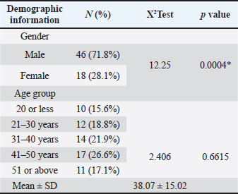
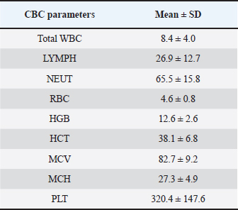
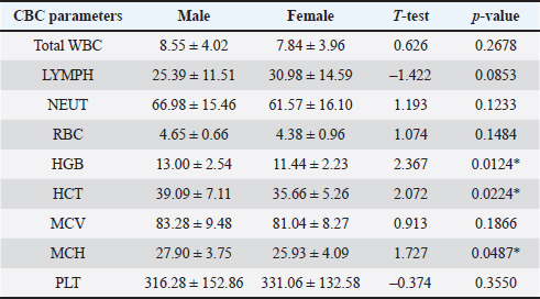
ABSTRACTBackground: Pulmonary tuberculosis (PTB) remains a major global health challenge, with significant morbidity and mortality. Beyond its respiratory manifestations, PTB is frequently associated with hematological alterations and elevated inflammatory markers, which may reflect disease severity and systemic inflammation. Data on these parameters among Libyan patients with PTB remain limited. Aim: This study aimed to evaluate complete blood count (CBC) parameters and inflammatory biomarkers, including the erythrocyte sedimentation rate (ESR) and C-reactive protein (CRP), in patients with PTB and to investigate the correlations between these inflammatory markers and CBC parameters. Methods: A retrospective cross-sectional study was conducted on 64 patients diagnosed with pulmonary TB between 2023 and 2025 at Al-Daran Hospital. Demographic data and laboratory results, including CBC and inflammatory markers (ESR and CRP), were extracted from medical archives. Statistical analysis included descriptive statistics, t-tests for group comparisons, and Pearson correlation to assess the relationships between inflammatory markers and CBC parameters. Results: Males comprised 71.8% of the study population, indicating a higher burden of PTB in men. CBC analysis demonstrated mild normocytic normochromic anemia, with a mean hemoglobin of 12.6 ± 2.6 g/dl and hematocrit of 38.1% ± 6.8%. Neutrophil percentages were slightly elevated (65.5% ± 15.8%), while lymphocyte counts remained within normal limits (26.9% ± 12.7%). Inflammatory markers were markedly elevated, with an ESR of 44.0 ± 38.4 mm/hour and a CRP level of 31.0 ± 48.1 mg/l, indicating active systemic inflammation. Females showed significantly lower hemoglobin, hematocrit, and MCH compared to males (p < 0.05). Correlation analysis revealed positive associations of CRP and ESR with total WBC count, neutrophils, and platelet count, and negative correlations with lymphocyte percentage and red blood cell indices (MCV and MCH), hemoglobin, and hematocrit. Conclusion: PTB is associated with mild normocytic normochromic anemia, neutrophilia, and elevated inflammatory markers. Although lymphocyte counts remained within normal ranges, they showed significant inverse correlations with inflammatory markers. Complete blood count and inflammatory parameters are strongly interrelated, highlighting their potential utility in assessing disease severity and monitoring patients with PTB. Keywords: Complete blood count, C-reactive protein, Erythrocyte sedimentation rate, Pulmonary tuberculosis. IntroductionPulmonary tuberculosis (PTB) is the most common and infectious form of tuberculosis, a contagious bacterial disease caused by Mycobacterium tuberculosis that continues to rank among the leading causes of infectious disease-related mortality worldwide (Ryan et al., 2025). The disease is primarily transmitted through airborne droplets expelled when individuals with active pulmonary infection cough or sneeze (Sarkar and Sarkar, 2025). Clinically, PTB is characterized by a persistent cough, chest pain, hemoptysis, fever, night sweats, weight loss, and generalized fatigue AlOsaimi et al., 2024). In 2023, tuberculosis affected an estimated 10.8 million people globally, reflecting its ongoing public health burden. In Libya, the incidence was estimated at 40 cases per 100,000 population, corresponding to nearly 2,800 new cases annually and indicating a moderate but persistent disease burden. Available regional data also suggest a higher prevalence among males, which has been attributed to occupational exposure, smoking habits, and differences in healthcare-seeking behavior (Organization, 2024; Lee et al., 2025). PTB is increasingly recognized as a systemic inflammatory condition that produces characteristic hematological changes beyond its respiratory manifestations. These alterations may provide useful adjuncts for disease assessment, particularly in settings where advanced diagnostic tools are limited (Batool et al., 2022; Asari et al., 2025). Reported abnormalities include anemia, changes in white blood cell counts (leukocytosis or leukopenia), neutrophilia, lymphocyte alterations, and platelet variations, along with elevated inflammatory markers such as erythrocyte sedimentation rate (ESR) and C-reactive protein (CRP) (Ştefanescu et al., 2021; Kumar et al., 2024). Among these, anemia remains the most frequently observed abnormality, affecting a substantial proportion of patients with active disease, with reported prevalence ranging from 30% to 85%, depending on population and disease severity (Dasaradhan et al., 2022; Tiu et al., 2025). The development of anemia in PTB is multifactorial. The most common mechanism is chronic anemia, characterized by impaired iron utilization, reduced erythropoietic activity, and altered iron storage profiles (Shah et al., 2022). Iron deficiency anemia may also coexist in some cases, particularly in populations affected by malnutrition (Begum and Latunde-Dada, 2019). At the molecular level, inflammatory cytokines, especially interleukin-6 (IL-6), stimulate hepatic hepcidin production, which reduces iron availability by trapping it within macrophages and limiting intestinal absorption, ultimately contributing to inflammation-associated anemia (Wrighting and Andrews, 2006; John and J, 2026). Several studies have supported these hematological patterns. Shah et al. (2022) reported decreased hemoglobin and hematocrit levels alongside elevated white blood cell counts and ESR in patients with PTB compared with healthy controls (Shah et al., 2022). Similar patterns have been observed in Eastern Sudan and Egypt, suggesting a consistent regional profile characterized by mild normocytic normochromic anemia, neutrophilia, and relative lymphocyte reduction in patients with pulmonary TB (Idriss et al., 2013; Ali et al., 2024). Although available data are limited and outdated in Libya, reports still indicate a male predominance and a notable burden of PTB, emphasizing the need for updated local evidence. Despite these findings, there remains a clear gap in current knowledge regarding the hematological and inflammatory profiles of patients with PTB in Libya, as most existing studies were conducted more than a decade ago and may not reflect current epidemiological or clinical patterns (Latifa Abdel-Hafid Jwieli and El-Majbri, 2020). A clearer understanding of these changes is important not only for improving diagnostic support but also for monitoring disease activity and guiding clinical management. Therefore, this study aimed to evaluate hematological alterations in patients with PTB at Al-Darnah Hospital, assess inflammatory biomarkers (ESR and CRP), and explore their correlations with complete blood count parameters to better characterize disease-related hematological and inflammatory changes. Materials and MethodsStudy design and settingThis retrospective cross-sectional analysis was conducted using previously documented medical records. Data were obtained from the medical archives of Al-Daran Hospital. Case records were reviewed over a 2-month period (October–November) and included patients diagnosed with PTB between 2023 and 2025. Study populationThe study population comprised 64 patients with microbiologically confirmed PTB. The diagnoses were established by the treating physicians at Al-Daran Hospital, following the institution’s routine diagnostic protocol. This protocol encompassed clinical presentation, radiological findings, and microbiological confirmation through AFB smear microscopy and/or the GeneXpert MTB/RIF assay. All eligible cases identified in the hospital records during the study period were included, without age- or sex-based selection. Inclusion was solely contingent on the availability of complete and clearly documented medical records. Information regarding the anti-TB treatment status of patients at the time of laboratory testing was not consistently available in the archived records and was therefore not assessed in the present study. Data collectionData were extracted from archived medical records, including patients’ demographic characteristics (age and sex) and complete blood count (CBC) parameters, such as hemoglobin (HGB), red blood cell (RBC) count, total white blood cell (WBC) count, lymphocyte and neutrophil percentages, hematocrit (HCT), platelet (PLT) count, mean corpuscular volume (MCV), mean corpuscular hemoglobin (MCH), and MCHC. Complete blood count analysis was performed using the Mission HA-360 Hematology Analyzer. Inflammatory biomarkers, including ESR and CRP, were also measured. ESR was measured using the Westergren method and reported in mm/hour, whereas CRP levels were determined using the Cobas Integra automated chemistry analyzer (Roche Diagnostics, Germany). To ensure data accuracy, only complete medical records were included in the analysis. All data were handled with strict confidentiality, and no personal identifiers were collected. The reference ranges for CBC parameters were based on the Practical Hematology of Dacie and Lewis 2006). Reference values for ESR and CRP were obtained from the Tietz Textbook of Clinical Chemistry and Molecular Diagnostics (Bruns et al., 2012). Statistical analysisData were analyzed using the appropriate statistical software. Continuous variables were expressed as mean ± SD, whereas categorical variables were expressed as frequencies and percentages. The independent sample t-test was used to assess differences between groups, and associations between variables were evaluated using Pearson’s correlation coefficient. The chi-square test was used to assess associations between categorical variables. A p-value of <0.05 was considered statistically significant. Ethical approvalThis retrospective study was conducted following an official request from the Faculty of Medical Technology, University of Tripoli, to the relevant authority at Al-Daran Hospital for permission to access archived medical records and collect data for research purposes. The hospital administration granted approval before data collection. This study involved only a retrospective review of archived records, with no direct patient contact and no collection of identifiable personal information. Therefore, informed consent was not required. Patient confidentiality and data privacy were strictly maintained throughout the study. ResultsDemographic characteristics of the study populationTable 1 presents the demographic characteristics of patients with PTB. A total of 64 patients were included during the study period (2023–2025). Males constituted the majority of cases (71.8%, n=46), whereas females accounted for 28.1% (n=18), with a statistically significant difference in gender distribution (χ²=12.25, p=0.0004). Patients were distributed across different age groups, with the highest proportion observed among those aged 41–50 years (26.6%), followed by 31–40 years (21.9%), 21–30 years (18.8%), ≥51 years (17.1%), and ≤20 years (15.6%). The mean age of the study population was 38.07 ± 15.02 years. No statistically significant difference was detected in age distribution (χ²=2.406, p=0.6615). Complete blood count parametersTable 2 summarizes the hematological profile of patients with pulmonary TB. The mean total WBC count was 8.4 ± 4.0 × 10⁹/l, which is within the reference range. The mean lymphocyte percentage was 26.9% ± 12.7%, which was also within normal limits, whereas the mean neutrophil percentage was 65.5% ± 15.8%, which slightly exceeded the normal range and showed a statistically significant difference (p=0.0429). The mean RBC count was 4.6 ± 0.8 × 10⁶ /µl and remained within normal limits. However, the mean HGB level was 12.6 ± 2.6 g/dl, and the mean HCT was 38.1% ± 6.8%, both of which were slightly below the standard reference range, with a statistically significant reduction in HCT (p=0.0427). RBC indices showed mean values of 82.7 ± 9.2 fL for MCV and 27.3 ± 4.9 pg for MCH, both within normal ranges. The mean PLT count was 320.4 ± 147.6 ×10⁹/l, which also falls within the reference range (Table 2). Gender-based comparison of the CBC parametersTable 3 illustrates the comparison of CBC parameters between male and female patients. No statistically significant differences were observed between genders with respect to total WBC count, lymphocyte percentage, neutrophil percentage, RBC count, MCV, or platelet count (p > 0.05). However, females exhibited significantly lower mean HGB levels (11.44 ± 2.23 g/dl) and HCT values (35.66% ± 5.26%) than males (13.00 ± 2.54 g/dl and 39.09% ± 7.11%, respectively), with statistically significant differences (p=0.0124 and p=0.0224). In addition, MCH was significantly lower in females (25.93 ± 4.09 pg) than in males (27.90 ± 3.75 pg) (p=0.0487). Table 1. Demographic information distribution among the samples.
Table 2. Mean and SD of the CBC parameters among the total samples.
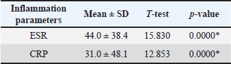
Inflammatory biomarkersTable 4 presents the inflammatory markers assessed in this study. The mean ESR was markedly elevated at 44.0 ± 38.4 mm/hour compared with the normal reference range. The mean CRP level was 31.0 ± 48.1 mg/l, which is considerably higher than the normal values. Both ESR and CRP levels were significantly elevated (p < 0.001). Table 3. Descriptive statistics of differences in CBC parameters between males and females.
Table 4. Mean and SD of inflammation parameters among the total samples.
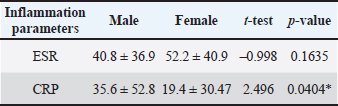
Table 5. Descriptive statistics of differences in inflammation parameters between males and females.
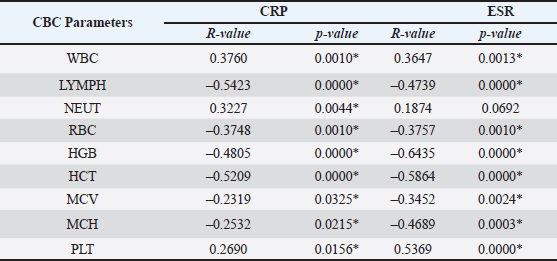
Gender-based comparison of inflammatory markersAs shown in Table 5, females demonstrated higher mean ESR values (52.2 ± 40.9 mm/hour) than males (40.8 ± 36.9 mm/hour); however, this difference was not statistically significant (p=0.1635). In contrast, males had significantly higher mean CRP levels (35.6 ± 52.8 mg/l) than females (19.4 ± 30.5 mg/l), with a statistically significant difference (p=0.0404). Correlation between inflammatory markers and CBC parametersPearson’s correlation analysis revealed significant associations between inflammatory markers and hematological parameters (Table 6). CRP levels were moderately positively correlated with total WBC count (r=0.3760, p=0.0010), neutrophil percentage (r=0.3227, p=0.0044), and PLT count (r=0.2690, p=0.0156). In contrast, CRP levels were strongly negatively correlated with lymphocyte percentage (r=−0.5423, p < 0.001) and red blood cell indices, including red blood cell count, hemoglobin, hematocrit, MCV, and MCH (p < 0.05). Similarly, ESR was positively correlated with total WBC count (r=0.3647, p=0.0013) and PLT count (r=0.5369, p < 0.001), while showing strong negative correlations with lymphocyte percentage and RBC parameters, particularly hemoglobin (r=−0.6435, p < 0.001) and hematocrit (r=−0.5864, p < 0.001). The correlation between ESR and the percentage of neutrophils was weak and not statistically significant (p=0.0692). DiscussionThis retrospective cross-sectional study evaluated hematological and inflammatory alterations among patients with pulmonary TB at Al-Daran Hospital. The findings demonstrate characteristic abnormalities in CBC parameters and markedly elevated inflammatory markers, with strong correlations between inflammation and hematological changes. Overall, the results are consistent with those of previous regional and international studies, although some variations were observed in the magnitude of the changes. Males constituted most pulmonary TB cases (71.8%) in the current study, indicating a pronounced male predominance. This finding is consistent with several international studies reporting higher prevalence of pulmonary TB among men. Batool et al. reported a male-to-female ratio of 2.5:1, while Mohammed et al. observed that 62.5% of patients with PTB were male (Mohammed, 2016; Batool et al., 2022). Similar trends have also been reported in regional data from Libya, where pulmonary TB notifications show a clear male predominance (Balakrishnan, 2022). The higher burden of pulmonary TB among males than females may be attributed to multiple factors, including increased occupational exposure, higher smoking rates, delayed healthcare-seeking behavior, and greater social mobility. These explanations have been widely suggested in epidemiological studies of PTB in low- and middle-income countries (Yang et al., 2024). Table 6. Pearson correlation coefficient between CRP/ESR and CBC parameters.
This study identified several hematological abnormalities associated with pulmonary TB. The mean hemoglobin (12.6 ± 2.6 g/dl) and hematocrit levels (38.1% ± 6.8%) were slightly below normal ranges, indicating mild anemia, predominantly of the normocytic normochromic type, as supported by normal MCV and MCH values. These findings agree with those of Shah et al, who reported significantly reduced hemoglobin and hematocrit levels in patients with PTB (Shah et al., 2022). Similar observations were reported in patients from Eastern Sudan by Idriss et al. (2013)and by Mohammed (2016) reinforcing the consistency of anemia as a common hematological manifestation of PTB (Idriss et al., 2013; Mohammed, 2016). The relatively mild degree of anemia observed in this study may reflect earlier diagnosis, less advanced disease, or variations in nutritional status and comorbidities among the study population (de Mendonça et al., 2021). Gender-based analysis revealed that females had significantly lower hemoglobin, hematocrit, and MCH values than males. This finding is consistent with previous studies and may be attributed to a higher susceptibility to iron deficiency among females, compounded by chronic inflammation associated with PTB (De Mendonça et al., 2021; Abaynew et al., 2023). The neutrophil percentage was slightly elevated (65.5% ± 15.8%), reflecting an active inflammatory response typical of pulmonary TB. This finding aligns with Shah et al, who reported significantly increased neutrophil counts in patients with PTB (Shah et al., 2022). In contrast, lymphocyte percentages in the present study remained within the normal range, although correlation analysis revealed significant negative associations with inflammatory markers (Omair et al., 2024). Differences in lymphocyte responses have been previously reported. For example, Ali et al. (2024) observed elevated lymphocyte counts in certain pulmonary TB populations, suggesting that lymphocyte dynamics may vary depending on disease severity, immune status, or drug-resistance patterns (Ali et al., 2024). These variations may explain the relatively preserved lymphocyte levels observed in the current study (Kulesh et al., 2025). Inflammatory biomarkers were significantly elevated in the current study, with a mean ESR of 44.0 ± 38.4 mm/h and a CRP level of 31.0 ± 48.1 mg/l, indicating active systemic inflammation. These results are consistent with those of previous studies by Shah et al. and Rohini et al., which also reported notably increased ESR and CRP levels in patients with pulmonary TB (Rohini et al., 2016; Shah et al., 2022). In addition, Batool et al. (2022) found elevated ESR in nearly all pulmonary TB cases (Batool et al., 2022). Gunluoglu et al. (2014) further demonstrated that ESR and CRP levels were significantly higher in patients with active pulmonary TB than in healthy controls (Gunluoglu et al., 2014). Together, these findings underscore the reliability of ESR and CRP as disease activity and inflammatory burden indicators in pulmonary TB. Correlation analysis revealed significant relationships between inflammatory markers and hematological parameters. CRP and ESR were positively correlated with total WBC count, neutrophils, and platelet count, indicating leukocytosis and reactive thrombocytosis in the presence of heightened inflammation (Tang et al., 2025). Similar correlations were reported by Batool et al. (2022) supporting the role of inflammatory cytokines in stimulating leukocyte and PLT production during active pulmonary TB (Batool et al., 2022). Conversely, lymphocyte counts were strongly negatively correlated with CRP and ESR, suggesting inflammation-associated lymphopenia. This observation is consistent with the findings of studies conducted in Egypt by Ali et al. (2024)and Rohini et al. (2016). In addition, negative correlations were observed between inflammatory markers and RBC indices, including HGB, HCT, MCV, and MCH. These findings indicate anemia related to chronic inflammation, potentially mediated by iron sequestration and impaired erythropoiesis, mechanisms commonly described in TB-associated anemia (John and J, 2026). Mohammed et al. and Idriss et al. reported similar inverse relationships between inflammatory markers and red cell parameters, further highlighting the impact of chronic inflammation on hematological profiles in patients with pulmonary TB (Idriss et al., 2013; Mohammed, 2016). Overall, the current study underscores the association of PTB with substantial hematological and inflammatory alterations, characterized by significant deviations in complete blood count parameters alongside elevated ESR and CRP levels. Although such associations are well-documented in international literature, empirical data within the Libyan context remain sparse. Consequently, this study offers locally derived evidence that enhances the regional understanding of TB-related laboratory abnormalities and establishes a critical baseline for future investigations in comparable clinical settings. This study has several limitations. Its retrospective cross-sectional design precludes the establishment of causal relationships. Additionally, the single-center setting and relatively small sample size may limit the generalizability of the findings. Although all patients were diagnosed according to the hospital’s routine diagnostic protocol using clinical, radiological, and microbiological criteria, the archived medical records did not consistently provide sufficient detail to permit subgroup analyses based on clinical or radiological findings. Information regarding anti-TB treatment status at the time of laboratory testing was also unavailable. Therefore, further prospective multicenter studies are warranted to validate and extend these findings. ConclusionThis study demonstrates that PTB is significantly associated with mild normocytic normochromic anemia, neutrophilia, and markedly elevated inflammatory markers. An inverse relationship was observed between lymphocyte counts and systemic inflammation markers. These findings suggest that routine hematological and inflammatory parameters may serve as valuable, cost-effective adjunctive tools for the clinical assessment and monitoring of patients with PTB, particularly in resource-limited settings. AcknowledgmentsThe authors sincerely thank the Al-Daran Hospital for their valuable support and assistance in data collection for this study. FundingNo external funding was received for this study Authors’ contributionsEman Abdulwahed conceived and developed the study concept, supervised the research, and contributed to the drafting and critical revision of the manuscript. Narjes Naser served as the co-supervisor and contributed to the study design, manuscript writing, editing, and final revision. Eman Abdullh, Esra Oweidat, and Boshra Altegazi were responsible for data collection and analysis. All authors have reviewed and approved the final version of the manuscript. Conflict of interestThe authors have no conflicts of interest to declare regarding the publication of this paper. Data availabilityAll data supporting this study’s findings are available within the manuscript. ReferencesAbaynew, Y., Ali, A., Taye, G. and Shenkut, M. 2023. Prevalence and types of anemia among people with tuberculosis in Africa: a systematic review and meta-analysis. Sci. Rep. 13, 5385. Ali, A., Wu, L., Moazen, E.M., Elsawy, S.B., Salama, K.S.M., Ullah, K., Elfeky, S.E.F., Alharbi, S.H. and Saleh, M.M. 2024. Hematological profiles and mortality risk in critically ill and drug-resistant tuberculosis patients: insights from a longitudinal study. Egypt. J. Bronchology. 18(1), 40. AlOsaimi, H.M., Alshammari, M.K., Almijlad, G.K., Alotaibi, N.M., Alqahtani, D.A., Alshamrani, M.M., Shutur, T.A., Alhazmi, M.F., Hurubi, M.A., ALShammari, K.S., Alzahrani, K.M., Aldaghriri, H.M., Alshammari, A.A., Alatawi, O.S. and Alharbi, R.A. 2024. Prevalence, clinical characteristics and determinants of unsuccessful treatment outcomes among pulmonary tuberculosis patients: a 5-year registry-based retrospective cohort study. Patient Related Outcome Measures 15, 187–198; doi: 10.2147/PROM.S463396 Asari, H.D., Berani, J.B., Patel, P.N. and Jehwani, D.P. 2025. A study of hematological profile in patient’s of tuberculosis. Eur. J. Cardiovasc. Med. 15(3). Balakrishnan, V.S. 2022. The burden of tuberculosis in Libya. Lancet. Respiratory Med. 10(11), e99–e100. Batool, Y., Pervaiz, G., Arooj, A. and Fatima, S. 2022. Hematological manifestations in patients newly diagnosed with pulmonary tuberculosis. Pak. J. Med. Sci. 38(7), 1968–1972; doi:10.12669/pjms.38.7.5911 Begum, S. and Latunde-Dada, G.O. 2019. Anemia of inflammation with an emphasis on chronic kidney disease. Nutrients 11(10), 11; doi:10.3390/nu11102424 Bruns, D.E., Tietz, N.W., Burtis, C.A. and Ashwood, E.R. 2012. Tietz textbook of clinical chemistry and molecular diagnostics, 5th ed. St. Louis, MI: Elsevier/Saunders. Dacie, J.V. 2006. Dacie and Lewis practical hematology, 10th ed. Philadelphia, PA: Churchill Livingstone Elsevier Health Sciences. Dasaradhan, T., Koneti, J., Kalluru, R., Gadde, S., Cherukuri, S.P. and Chikatimalla, R. 2022. Tuberculosis-associated anemia: a narrative review. Cureus 14(8), e27746; doi:10.7759/cureus.27746 De Mendonça, E.B., Schmaltz, C.A., Sant’Anna, F.M., Vizzoni, A.G., Mendes-De-Almeida, D.P., De Oliveira, R.D.V.C. and Rolla, V.C. 2021. Anemia in tuberculosis cases: a biomarker of severity?. PLos One. 16(2), 245458. Gunluoglu, G., Yazar, E.E., Veske, N.S., Seyhan, E.C. and Altin, S. 2014. Mean platelet volume as an inflammation marker in active pulmonary tuberculosis. Multidiscip. Respir. Med. 9(1), 11; doi:10.1186/2049-6958-9-11 Idriss, M.I., Khalil, H.B., Abbashar, A.O., Widaah, S. and Magzoub, M. 2013. Hematological changes among tuberculous Sudanese patients in Kassala Area, Eastern Sudan. J. Intensive Care Med. . John, J. 2026. Association between hepcidin–ferroportin axis dysregulation and disease severity in pulmonary tuberculosis Kulesh, V., Peskov, K., Helmlinger, G. and Bocharov, G. 2025. Systematic review and quantitative meta-analysis of age-dependent human T-lymphocyte homeostasis. Front. Immunol. 16, 1475871; doi:10.1016/j.fimmu.2025.0871 Kumar, S.R., Kandhasamy, C., Velayutham, V.B., Chinnaiyan, P., Kannan, M., Jawahar, M.S. and Padmapriyadarsini, C. 2024. Hematological parameters in patients with pulmonary tuberculosis and its presentation among favorable and unfavorable treatment outcomes. Indian J. Public Health 68(3), 362–365; doi:10.4103/ijph.ijph_918_23 Latifa Abdel-Hafid Jwieli, H. A. E.-M. and El-Majbri, H. G. (2020). Survey of tuberculosis patient cases in Quefia Chest Hospital in Benghazi, Libya between 2010 and 2018. Global Scientific, 8(3), 14. Lee, H., Kim, J., Kim, J., Park, Y. and -J. 2025. Review of the global burden of tuberculosis in 2023: insights from the World Health Organization Global Tuberculosis Report 2024. Public Health Weekly Rep. 18(11), 55. Mohammed, M.A. 2016. Some hematological parameters among patients with pulmonary tuberculosis–Khartoum state. J. Clin. Oncol. Omair, M., Baig, M.S., Farooqui, W.A., Kousar, S., Noori, M.Y., Zeehan, N., Khan, A., Isa, S., Kamran, D.S., Bari, M.F. and Mehmood, M. 2024. Relationship of neutrophil lymphocyte ratio, monocyte lymphocyte ratio and neutrophil monocyte ratio with treatment response in pulmonary tuberculosis patients during intensive phase treatment. BMC. Infect. Dis. 24(1), 1–7. Organization, W. H. 2024. Implementing the global health sector strategies on HIV, viral hepatitis and sexually transmitted infections, 2022–2030: report on progress and gaps 2024. Geneva: World Health Organization. https://doi.org/10.1016/j.who.2024.04.006. Rohini, K., Surekha Bhat, M., Srikumar, P.S. and Mahesh Kumar, A. 2016. Assessment of hematological parameters in pulmonary tuberculosis patients. Indian J. Clin. Biochem. 31(3), 332–335; doi:10.1007/s12291-015-0535-8 Ryan, J.M., Shelton, K., Dzieciatkowska, M., Kruh-Garcia, N. and Dobos, K.M. 2025. Establishment of minimum protein standards for Mycobacterium tuberculosis-derived extracellular vesicles through comparison of EV enrichment methods. Mycobacteria 1(1), 3; doi:10.1016/j.mycobacteria.2025.01.03 Sarkar, M. and Sarkar, J. 2025. Transmission of Mycobacterium tuberculosis. J. Assoc. Physicians. India. 73(9), 91–96; doi:10.59556/japi.73.1113 Shah, A.R., Desai, K.N. and Maru, A.M. 2022. Evaluation of hematological parameters in pulmonary tuberculosis patients. J. Fam. Med. Primary Care 11(8), 4424–4428; doi:10.4103/jfmpc.jfmpc_2451_21 Ştefanescu, S., Cocoş, R., Turcu-Stiolica, A., Mahler, B., Meca, A.D., Giura, A.M.C., Bogdan, M., Shelby, E.S., Zamfirescu, G. and Pisoschi, C.G. 2021. Evaluation of prognostic significance of hematological profiles after the intensive phase treatment in pulmonary tuberculosis patients from Romania. PLos One. 16(4), 249301; doi:10.1371/journal.pone.0249301 Tiu, D.N., Sirajuddin Ahmed Siddiqi, S.M.F.M. and Ranjan, R. 2025. Hematological changes in pulmonary tuberculosis: focus on anemia, disease severity, and therapeutic implications. Cureus 17(6), e86550; doi:10.7759/cureus.86550 Wang G Tang., Wang, C., Xu, R., Shen, C. and Li, G. 2025. Inflammatory markers CRP and WBC as predictors of liver function impairment in multiple injury trauma patients: a repeated measures analysis. Front. Med. 12, 1570474. Wrighting, D.M. and Andrews, N.C. 2006. Interleukin-6 induces hepcidin expression through STAT3. Blood 108(9), 3204–3209. Yang, H., Ruan, X., Li, W., Xiong, J. and Zheng, Y. 2024. Global, regional, and national burden of tuberculosis and attributable risk factors for 204 countries and territories, 1990–2021: a systematic analysis for the Global Burden of Diseases 2021 study. BMC Public Health, 24(1), 3111. | ||
| How to Cite this Article |
| Pubmed Style Abdulwahed E, Naser N, Abdullh E, Oweidat E, Altegazi B. Evaluation of complete blood count and inflammatory markers in pulmonary tuberculosis: A retrospective cross-sectional study at Al-Daran hospital. J Microbiol Infect Dis. 2026; 16(3): 172-178. doi:10.5455/JMID.2026.v16.i3.7 Web Style Abdulwahed E, Naser N, Abdullh E, Oweidat E, Altegazi B. Evaluation of complete blood count and inflammatory markers in pulmonary tuberculosis: A retrospective cross-sectional study at Al-Daran hospital. https://www.jmidonline.org/?mno=317065 [Access: August 28, 2026]. doi:10.5455/JMID.2026.v16.i3.7 AMA (American Medical Association) Style Abdulwahed E, Naser N, Abdullh E, Oweidat E, Altegazi B. Evaluation of complete blood count and inflammatory markers in pulmonary tuberculosis: A retrospective cross-sectional study at Al-Daran hospital. J Microbiol Infect Dis. 2026; 16(3): 172-178. doi:10.5455/JMID.2026.v16.i3.7 Vancouver/ICMJE Style Abdulwahed E, Naser N, Abdullh E, Oweidat E, Altegazi B. Evaluation of complete blood count and inflammatory markers in pulmonary tuberculosis: A retrospective cross-sectional study at Al-Daran hospital. J Microbiol Infect Dis. (2026), [cited August 28, 2026]; 16(3): 172-178. doi:10.5455/JMID.2026.v16.i3.7 Harvard Style Abdulwahed, E., Naser, . N., Abdullh, . E., Oweidat, . E. & Altegazi, . B. (2026) Evaluation of complete blood count and inflammatory markers in pulmonary tuberculosis: A retrospective cross-sectional study at Al-Daran hospital. J Microbiol Infect Dis, 16 (3), 172-178. doi:10.5455/JMID.2026.v16.i3.7 Turabian Style Abdulwahed, Eman, Narjes Naser, Eman Abdullh, Esra Oweidat, and Boshra Altegazi. 2026. Evaluation of complete blood count and inflammatory markers in pulmonary tuberculosis: A retrospective cross-sectional study at Al-Daran hospital. Journal of Microbiology and Infectious Diseases, 16 (3), 172-178. doi:10.5455/JMID.2026.v16.i3.7 Chicago Style Abdulwahed, Eman, Narjes Naser, Eman Abdullh, Esra Oweidat, and Boshra Altegazi. "Evaluation of complete blood count and inflammatory markers in pulmonary tuberculosis: A retrospective cross-sectional study at Al-Daran hospital." Journal of Microbiology and Infectious Diseases 16 (2026), 172-178. doi:10.5455/JMID.2026.v16.i3.7 MLA (The Modern Language Association) Style Abdulwahed, Eman, Narjes Naser, Eman Abdullh, Esra Oweidat, and Boshra Altegazi. "Evaluation of complete blood count and inflammatory markers in pulmonary tuberculosis: A retrospective cross-sectional study at Al-Daran hospital." Journal of Microbiology and Infectious Diseases 16.3 (2026), 172-178. Print. doi:10.5455/JMID.2026.v16.i3.7 APA (American Psychological Association) Style Abdulwahed, E., Naser, . N., Abdullh, . E., Oweidat, . E. & Altegazi, . B. (2026) Evaluation of complete blood count and inflammatory markers in pulmonary tuberculosis: A retrospective cross-sectional study at Al-Daran hospital. Journal of Microbiology and Infectious Diseases, 16 (3), 172-178. doi:10.5455/JMID.2026.v16.i3.7 |


