






| Research Article | ||
J Microbiol Infect Dis. 2026; 16(3): 137-144 J. Microbiol. Infect. Dis., (2026), Vol. 16(3): 137–144 Research Article Characterization of Candida species isolated from various clinical specimens in Misurata, Libya: Distribution and antifungal susceptibilityZaineb A. Elmahjoub1*, Ebrahim M. Daghman1 and Abdul-Rauf M. Khallil21Department of Microbiology, Faculty of Science, Misurata University, Misurata, Libya 2Department of Microbiology and Botany, Assiut University, Assuit, Egypt *Corresponding Author: Zaineb A. Elmahjoub. Department of Microbiology, Faculty of Science, Misurata University, Misurata, Libya. Email: zainebelmahjoub [at] gmail.com Submitted: 02/03/2026 Revised: 22/05/2026 Accepted: 03/06/2026 Published: 07/07/2026 © 2025 Journal of Microbiology and Infectious Diseases
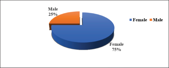
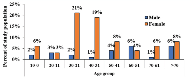
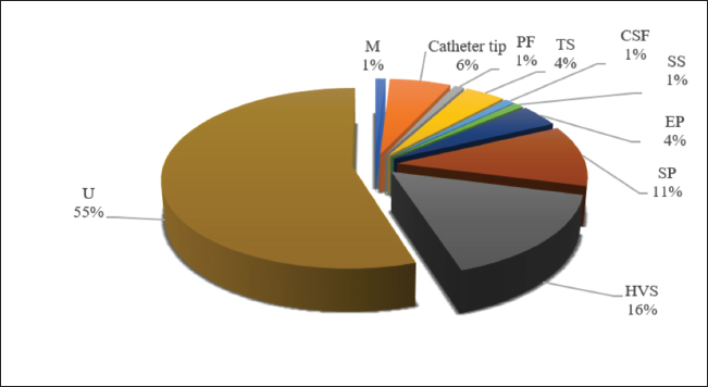
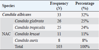
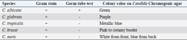
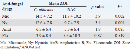
ABSTRACTBackground: Infections caused by Candida species, including Candida albicans and non-albicans Candida (NAC), have emerged as a significant global health challenge. This trend is driven by the increasing prevalence of immunocompromised populations and the rise of antifungal resistance, which complicates clinical management and increases morbidity. Aim: This study aimed to investigate the distribution of Candida spp. and evaluate their antifungal susceptibility profiles among patients in Misurata, Libya, to provide evidence-based data for optimizing therapeutic strategies. Methods: A prospective cross-sectional study was conducted between 2024 and 2025, analyzing 103 Candida-positive isolates from seven laboratories in the Misurata region. Identification was performed using Gram staining, culture on Sabouraud’s dextrose agar, yeast extract peptone dextrose, and chromogenic Candida agar. Antifungal susceptibility testing followed CLSI M44-A guidelines, using the disc diffusion method for Amphotericin B, Miconazole, Fluconazole, and Nystatin. Results: The study population comprised 25% males and 75% females, with the highest prevalence observed in the 21–40-years age group. NAC species predominated (68%), with Candida glabrata being the most frequent (25%), followed by Candida tropicalis (24%), Candida krusei (11%), and Candida auris (8%). Candida albicans accounted for 32% of isolates. Mixed infections were noted in three cases [C. tropicalis with C. albicans (67%) and C. tropicalis with C. krusei (33%)]. Urine specimens showed the highest prevalence of infection (55%), followed by high vaginal swabs (16%) and sputum (11%). Miconazole and Nystatin demonstrated the highest antifungal activity, while Fluconazole and Amphotericin B were less effective, particularly against NAC spp. Conclusion: Candidiasis represents a significant opportunistic infection in Misurata, with NAC species exhibiting increased resistance. Rapid identification using chromogenic media facilitates timely species detection, and ongoing antifungal susceptibility surveillance is essential for guiding effective therapy and limiting the spread of resistance. Keywords: Antifungal resistance, C. albicans, Candidiasis, Non-albicans Candida, Species distribution. IntroductionCandidiasis, the most common fungal infection affecting humans, is caused by Candida spp., which are endogenous organisms that can lead to opportunistic infections. The clinical manifestations of candidiasis are highly varied, encompassing acute, subacute, chronic, and episodic cases (Brown et al., 2012). The severity of these infections ranges from mild, self-limiting conditions to severe, potentially life-threatening illnesses. In recent years, the prevalence of certain predisposing factors has risen, contributing to a global increase in candidiasis cases (Brown et al., 2012; Pristov and Ghannoum, 2019; Lamoth et al., 2021). Effective identification of Candida spp. is crucial for appropriate management, especially as infections caused by non-albicans Candida (NAC) spp. have become more common. This growing prominence of NAC spp. has coincided with a global rise in candidiasis cases (Turner and Butler, 2014; Pappas et al., 2016). While Candida albicans continues to be the predominant cause of yeast infections, accounting for over half of all cases globally (Arendrup and Patterson, 2017), infections due to NAC spp. have shown an alarming upward trend. Notably, these species often demonstrate reduced sensitivity to antifungal therapies (Pfaller et al., 2019). In Libya, and specifically in the Misurata region, the epidemiological landscape of candidiasis requires updated data to address the challenges of antifungal stewardship. Effective patient management relies on rapid identification and accurate susceptibility profiling; yet many clinical settings still rely on empirical treatments that may exacerbate resistance (Turner and Butler, 2014; Arendrup and Patterson, 2017). Despite the global rise in NAC infections, there is a paucity of localized data regarding the specific distribution and resistance patterns of Candida spp. in Misurata, Libya. Most existing studies in the Misurata region are either outdated or do not provide a comprehensive analysis of both phenotypic identification and antifungal susceptibility testing across diverse clinical specimens. Therefore, this study addresses this gap by providing a contemporary characterization of Candida spp., including the emerging multidrug-resistant Candida auris, within the local clinical context, thereby providing data to support effective therapeutic strategies. Materials and MethodsStudy design and settingA prospective cross-sectional study was conducted between 2024 and 2025. The study involved the collection and analysis of 103 non-duplicate Candida isolates obtained from seven microbiology laboratories in Misurata, Libya. These laboratories serve a diverse patient population across various clinical departments. All tests were performed in the Microbiology Laboratory at Misurata Medical Center Clinical specimens collectionSpecimens were initially detected on blood agar plates, where Candida appeared as dry, creamy white, opaque colonies. Isolates were recovered from various clinical sources, such as urine, high vaginal swabs, sputum, and others, from patients across the departments of Pediatrics (3%), Maxillary facial (3%), Gynecology (16%), Surgery (1%), Medicine (7%), Dermatology (1%), Intensive Care Unite (30%), and Outpatient Clinics, (39%). Samples were gathered, and patient demographic details, such as age and gender, were recorded, along with origin of the specimens and the isolation date from the laboratory information system. Identification of Candida spp.A multi-step phenotypic approach was used for species-level identification: 1. Cultural examinationCandida spp. were subcultured on Sabouraud's dextrose agar media (Oxoid, UK) and yeast extract peptone dextrose agar media (Oxoid, UK), both supplemented with 0.05% chloramphenicol to inhibit bacterial growth. Plates were incubated at 37°C for 24–48 hours. 2. Microscopic examinationThe growth obtained from Sabouraud's dextrose agar media and yeast extract peptone dextrose agar media was subjected to Gram staining under microscopic examination according to a standard technique using crystal violet iodine complex, safranin, and alcohol to identify budding yeast cells and pseudohyphae. The preparation was examined using the X10 and X40 objective lenses. 3. Germ tube formationA rapid screen to differentiate C. albicans from NAC spp. A tiny piece of isolated colonies was transferred using a sterile stick and mixed with 0.5 ml of human serum, then incubated at 37°C for 3 hours. A drop of the serum-containing yeast culture was placed on a clean microscope glass slide and covered with a cover glass. The preparation was examined using X10 and X40 objective lenses. Sprouting yeast cells (tube-like outgrowths), also known as germ tubes, were observed in C. albicans. 4. Differential identificationIsolates were subcultured on Candida chromogenic agar plates (BioMérieux, UK) and incubated at 37°C for 24–48 hours. Species were presumptively identified based on distinct colony colors: C. albicans: green, Candida glabrata: purple, Candida tropicalis: metallic blue, Candida krusei: pink with creamy borders, and C. auris: white (front)/blue (back). If there are mixed species, we observed more than one color on the plate. In this case, we should repeat the culture for each colony in a single plate to separate the species from each other. Antifungal susceptibility testingAll isolates were tested using the disc diffusion method with most routine antifungal discs, including Amphotericin B (100 IU), Fluconazole (25 μg), Miconazole (10 μg), and Nystatin (100 IU) (Liofilchem, Italy). Before starting the analysis, test strains from long-term stock cultures were subcultured on yeast extract peptone dextrose and incubated at 37°C for 48 hours. The following day, Candida colonies were harvested from subcultures and suspended in 2 ml of sterile saline. The turbidity was standardized to 0.5 McFarland (Remel, USA) before the suspension was spread across yeast extract peptone dextrose plates (Oxoid, UK). Antifungal discs were then placed on the plates, which were incubated again at 37°C for 48 hours. After incubation, the diameters of inhibition zones (ZOI) around the antifungal discs were measured in millimeters and interpreted according to the clinical and laboratory standards institute 2010 guidelines (CLSI M44-A). Statistical analysisData were managed using Microsoft Excel and analyzed via Statistical Package for the Social Sciences. ANOVA test was utilized to compare the mean ZOI between C. albicans and NAC spp. A p-value <0.05 was defined as statistically significant. Ethical approvalNot needed for this study. However, before sample collection, the Head of the Microbiology Department provided a letter of introduction that was submitted to the Central Library of the Libyan Authority for Scientific Research for formal approval. Next, formal permission was obtained from the heads of the laboratories. ResultsThe study examined the distribution of Candida infections in relation to patient age and gender, identifying various Candida spp. isolated from different clinical samples during the study period, as shown in the Figures 1–3 and Tables 1–2, while Table 3 shows the results of identification tests of Candida spp. The study also evaluated the efficacy of antifungal drugs, as shown in Table 4. Table 5 shows that Miconazole and Nystatin exhibited a significant difference in their effects between CA and NAC (p-value <0.05), whereas Amphotericin B and Fluconazole displayed contrary effects across the CA and NAC (p-value >0.05), as determined by the ANOVA test. DiscussionCandidiasis, an infection caused by members of the Candida genus, can manifest as either a primary or secondary condition. These infections can range from localized to systemic (Offianan et al., 2016). Over the last two decades, the epidemiology of Candida infections has shifted significantly, with a marked increase in the number of affected individuals (Willinger and Manafi, 1999). Both C. albicans and NAC spp. are recognized as opportunistic pathogens capable of causing severe systemic infections, particularly among immunocompromised patients in intensive care settings (Willinger and Manafi, 1999). In the present study, a notable gender disparity was observed, with a higher prevalence of infection among females (75%) compared to males (25%). This finding aligns with the trend reported by authors who also found a female predominance (60.2% vs. 39.8%) (Amar et al., 2013). However, it contrasts with the results of Athokpam et al. (2022) where a higher proportion of infections was recorded in males (57%) than in females (43%) (Athokpam et al., 2022).
Fig. 1. Distribution of Candida spp. by the gender of patients.
Fig. 2. Age- and gender-wise distribution of Candida spp.
Fig. 3. Distribution of anatomical sites of clinical samples. TS: throat swab, HVS: high vaginal swab, U: urine, SP: sputum, CSF: cerebral spinal fluid, PF: pleural fluid, EP: ear pus, M: mini-Bal, ETA: endotracheal aspirate, SS: skin scrap. Table 1. Distribution of Candida spp.
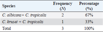
Table 2. Distribution of mixed infections.
The variation in these proportions across different studies may be attributed to differences in sample size, regional demographics, or specific underlying risk factors within the study populations. The predominance in females may be related to factors such as urinary tract infections, vaginitis, and prolonged exposure to water often associated with housework (Loster et al., 2016; Athokpam et al., 2022). In this assessment, 103 Candida spp. were examined, derived from clinical specimens across age groups ranging from a 1-year-old infant to an 87-year-old adult. The highest percentage of Candida spp. was observed in the females with the 21–30 years age group, comprising 21%, of cases, followed by the 31–40 years group at 19%. The lowest percentage was noted among individuals aged 11–20 years, at just 3%. These findings are consistent with earlier research identifying the age group of 21–40 years as most susceptible to Candida spp. (Patel et al., 2012; Raveendran and Guru, 2016). Factors such as hormonal fluctuations, heightened sexual activity, and higher pregnancy rates within this age group may contribute to this trend. This study found most Candida spp. in urine samples (55%), followed by high vaginal swabs (16%), sputum (11%), catheter tips (6%), and 4% each from ear and oral swabs. Only 1% were recovered from skin scrapings, pleural fluid, cerebrospinal fluid, and mini-bronchoalveolar lavage (mini-Bal). Previous studies also reported a high proportion of isolates from urine samples, such as 46.9% and sputum (28.78%) (Joseph et al., 2017). Similarly, another study found the majority of isolates in urine samples (60%), followed by respiratory specimens (17.3%) (Shaik et al., 2016). Conversely, one study reported sputum samples as the leading source of Candida isolates (43%), followed by urine (34%), blood (7%), nail clippings (6%), oral swabs (5%), vaginal swabs (2%), with 1% each from skin scrapings, catheter tips, and pus from diabetic foot infections (Athokpam et al., 2022). This may be attributed to the increasing prevalence of fungi, including both yeasts and molds, in sputum samples. Another study focused on Candida isolates from high vaginal swabs, reporting a distribution of 50.5% high vaginal swabs, 25% urine, 15.7% cervical swabs, and 8.8% ear swabs (Guru, 2016). Table 3. Identification tests of Candida spp.
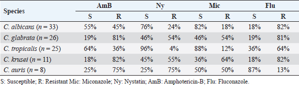
Table 4. Percent efficacy of antifungal drugs against Candida spp.
Table 5. Comparison of ZOI of antifungal drugs among Candida spp.
Overall, Candida spp. is considered one of the most common opportunistic pathogens and ranks as the fourth leading cause of hospital-acquired infection among immunocompromised patients globally over the past two decades (Al Thaqafi et al., 2014; Yamin et al., 2021). This study underscores the dominance of NAC spp. over C. albicans, with NAC accounting for 68% of the total isolates, while C. albicans comprised 32%. Mixed isolates represented only three cases. Among NAC spp. C. glabrata emerged as the most prevalent (25%), followed by C. tropicalis (24%), C. krusei (11%), and C. auris (8%). Mixed isolates were C. tropicalis with C. albicans (2/3(67%)) and C. tropicalis with C. krusei) (1/3 (33% )). These findings are consistent with a prior study where 47.7% of isolates were identified as C. albicans, while 52.3% belonged to NAC spp. Among the NAC group, Candida parapsilosis accounted for 36.9%, followed by C. krusei (5.4%), C. tropicalis (2.7%), C. glabrata (2.7%), and a combined 4.5% encompassing C. kefyr, C. famata, and C. lusitaniae (Eksi et al., 2013). Another study reported C. albicans as the dominant species at 18%, with C. dubliniensis representing 8%, C. glabrata 13%, and C. tropicalis 1% (Nurat et al., 2016). The antifungal susceptibility profile of C. albicans in this study revealed varying degrees of effectiveness across the tested antifungal agents. Miconazole demonstrated the highest efficacy, with 82% of isolates showing high susceptibility. This strong response aligns with the findings of a previous study, which reported Miconazole as having a significant fungicidal effect against C. albicans (Vandenbosch et al., 2010). Nystatin followed closely with a significant sensitivity rate of 76%. This level of effectiveness of Nystatin on C. albicans is notably higher than the 63.5% reported in a previous study (Khan and Baqai, 2010). In contrast, Amphotericin B exhibited the lowest sensitivity (55%) among the effective drugs against C. albicans, consistent with observations by authors whose work indicated that C. albicans may show reduced sensitivity to Amphotericin B compared to other antifungal agents (Sinh et al., 2021). Most notably, a high level of resistance was observed toward Fluconazole, with 82% of isolates proving resistant. This indicates a significantly higher resistance pattern than the 36.2% reported in a previous study (Fan et al., 2023). NAC spp. in this study presented even more formidable therapeutic challenges, often displaying multidrug-resistant phenotypes. The antifungal susceptibility profile of C. glabrata observed in this study presents a critical therapeutic challenge, characterized by a low sensitivity of only 19% to both Fluconazole and Amphotericin B. The high resistance to Fluconazole is largely attributed to the species' haploid nature, which facilitates the rapid expression of resistance mutations (Pfaller et al., 2022). Furthermore, while C. glabrata has historically shown higher susceptibility to polyenes, the 19% sensitivity to Amphotericin B found here suggests the emergence of Multidrug resistant (MDR) C. glabrata strains, likely driven by genetic alterations or mitochondrial dysfunction that reduces ergosterol levels in the cell membrane, as shown in a recent study (Eliaš et al., 2024). Although Miconazole and Nystatin performed comparatively better, their moderate sensitivity rate of 46% still indicates significant cross-resistance within these isolates (Parums, 2022). The antifungal susceptibility profile of C. tropicalis in this study reinforces the species' reputation for developing significant resistance while maintaining sensitivity to certain polyenes and imidazoles. The high sensitivity to Nystatin (96%) and Miconazole (88%) suggests these remain potent options for local or systemic applications. However, the moderate response to Amphotericin B (64%) and the alarmingly low sensitivity to Fluconazole (36%) signal a major shift in the treatment landscape for this pathogen. Recent studies confirm that C. tropicalis is no longer a predictably sensitive species. The rise in Fluconazole resistance is largely driven by genetic mutations and the overexpression of efflux pumps, which are frequently selected for in hospital environments (Arastehfar et al., 2020; Fan et al., 2023). This makes C. tropicalis a primary contributor to the growing burden of NAC infections globally. The antifungal susceptibility profile of C. krusei in this study confirms its intrinsic resistance to Fluconazole (18%) reveals a similarly low sensitivity to Amphotericin B (18%) and Miconazole (36%). In contrast, Nystatin demonstrated better efficacy, with a 45% sensitivity rate. The resistance pattern aligns with established clinical literature, which identifies C. krusei as a naturally resistant pathogen, thereby underscoring the critical necessity of species-level identification to ensure appropriate antifungal therapy (Pfaller et al., 2010; Arendrup, 2014; Pappas et al., 2018). The antifungal susceptibility profile of C. auris in this study reinforces its status as a significant MDR pathogen, exhibiting a critical lack of sensitivity to Fluconazole (13%), Amphotericin B (25%), and Nystatin (25%), and Miconazole (50%). These findings are consistent with global surveillance data identifying C. auris as an emerging public health threat characterized by healthcare-associated outbreaks and highly restricted therapeutic options (Chowdhary et al., 2016; Berkow and Lockhart, 2017). Broadly, Miconazole and Nystatin demonstrated the highest efficacy across the Candida genus in this study. Conversely, Fluconazole and Amphotericin B showed diminished effectiveness, a trend especially pronounced in NAC spp. This shift in susceptibility patterns reflects contemporary global antifungal resistance trends, emphasizing the clinical necessity of routine antifungal susceptibility testing to ensure targeted patient management and robust antifungal stewardship (Pappas et al., 2018; Arastehfar et al., 2020; Parums, 2022; Cornely et al., 2025). The observed resistance patterns, especially for Fluconazole, underscore the importance of species-level identification and individualized susceptibility testing to guide appropriate therapeutic interventions. The varying efficacy of antifungal agents across different Candida spp emphasizes the need for continuous surveillance of antifungal resistance trends to optimize patient outcomes. ConclusionThis study concludes that Candida spp., particularly C. albicans and NAC, are critical opportunistic pathogens predominantly affecting immunocompromised individuals, especially females aged 21–40 years. NAC spp. are the predominant cause of candidiasis in Misurata, Libya, and exhibit alarming levels of resistance to first-line antifungal agents like fluconazole. The use of Candida chromogenic media has proven to be an effective and rapid tool for presumptive identification. However, the high resistance rates observed emphasize that antifungal treatment should be guided by laboratory-confirmed susceptibility testing rather than empirical protocols. Future research should focus on the molecular mechanisms of resistance and longitudinal surveillance to monitor the spread of multidrug-resistant strains like C. auris. AcknowledgmentsWe especially thank and acknowledge my supervisors and the management of the Faculty of Science, Misurata University, for their invaluable support throughout this study. We also thank the entire staff of the microbiology laboratories in the Misurata region for their support and assistance. Conflict of interestThe authors declare that the research was conducted in the absence of any commercial or financial relationships that could be construed as a potential conflict of interest. FundingAll authors have read and agreed to the published version of the article. Authors’ contributionsZaineb A. Elmahjoub was responsible for collecting clinical samples, performing experiments, analyzing data, and writing the original draft of the article. Ebrahim M. Daghman and Abdul-Rauf M. Khallil supervised, reviewed, and edited the article. Data availabilityAll data presented in this study are provided within the manuscript. ReferencesAl Thaqafi, A.H.O., Farahat, F.M., Al Harbi, M.I., Al Amri, A.F.W. and Perfect, J.R. 2014. Predictors and outcomes of Candida bloodstream infection: eight-year surveillance, western Saudi Arabia. Int. J. Infect. Dis. 21(5), 5–9. Amar, C., Ashish, J., Vinay Hajare, V.H., Sreekantha, S., Yogesh, B. and Vinodchandran, V. 2013. Study of prevalence and antifungal susceptibility of Candida. Int. J. Pharma. Bio. Sci. 4(2), 361–381. Arastehfar, A., Gabaldón, T., Garcia-Rubio, R., Jenks, J.D., Hoenigl, M., Salzer, H.J.F., Ilkit, M., Lass-Flörl, C. and Perlin, D.S. 2020. Drug-resistant fungi: an emerging challenge threatening our limited antifungal armamentarium. Antibiotics 9(12), 877. Arendrup, M. 2014. Update on antifungal resistance in Aspergillus and Candida. Clinical microbiology and infection, 20, 42-48. J. Infect. Dis. 216 (suppl_3), S445–S451. Arendrup, M.C. and Patterson, T.F. 2017. Multidrug-resistant Candida: epidemiology, molecular mechanisms, and treatment. J. Infect. Dis. 216(suppl_3), S445–S451. Athokpam, D.C., Haobam, S., Singh, K.M. and Chongtham, U. 2022. Speciation of Candida isolates from various clinical specimens in a tertiary care hospital, Manipur. J. Cardiovasc. Dis. Res. 13(1), 412–422. Berkow, E.L. and Lockhart, S.R. 2017. Fluconazole resistance in Candida species: a current perspective. Infect. Drug. Resist. 10, 237–245. Brown, G.D., Denning, D.W., Gow, N.A.R., Levitz, S.M., Netea, M.G. and White, T.C. 2012. Hidden killers: human fungal infections. Sci. Translational. Med. 4(165), 165rv113. Chowdhary, A., Voss, A. and Meis, J.F. 2016. Multidrug-resistant Candida auris: ‘new kid on the block’ in hospital-associated infections?. J. Hosp. Infection. 94(3), 209–212. Cornely, O.A., Sprute, R., Bassetti, M., Chen, S.C., Groll, A.H., Kurzai, O., Lass-Flörl, C., Ostrosky-Zeichner, L., Rautemaa-Richardson, R. and Revathi, G. 2025. Global guideline for the diagnosis and management of candidiasis: an initiative of the ECMM in cooperation with ISHAM and ASM. Lancet Infect. Dis. 25, e280–93. Eksi, F., Gayyurhan, E.D. and Balci, I. 2013. In vitro susceptibility of Candida species to four antifungal agents assessed by the reference broth microdilution method. Scientific. World. J. 1(1), 236903. Eliaš, D., Tóth Hervay, N. and Gbelská, Y. 2024. Ergosterol biosynthesis and regulation impact the antifungal resistance and virulence of Candida spp. stresses. Stresses 4(4), 641–662. Fan, X., Tsui, C.K.M., Chen, X., Wang, P., Liu, Z.J. and Yang, C.X. 2023. High prevalence of fluconazole-resistant Candida tropicalis among candiduria samples in China: an ignored matter of concern. Front. Microbiol. 14, 1125241. Joseph, K., Ameena, K.K. and George, A.T. 2017. A study on proportion, speciation and antifungal resistance pattern of the Candida isolates in a Tertiary Care Hospital of North Kerala, India. Int. J. Curr. Microbiol. App. Sci. 6(5), 434–439. Khan, F. and Baqai, R. 2010. In vitro antifungal sensitivity of fluconazole, clotrimazole and nystatin against vaginal candidiasis in females of childbearing age. J. Ayub Med. Coll. Abbottabad 22(4), 197–200. Lamoth, F., Lewis, R. and Kontoyiannis, D. 2021. Role and interpretation of antifungal susceptibility testing for the management of invasive fungal infections. J. Fungi 7, 17. Loster, J.E., Wieczorek, A. and Loster, B.W. 2016. Correlation between age and gender in Candida species infections of complete denture wearers: a retrospective analysis. Clin. Interventions. Aging. 1707, 1707–1714. Nurat, A., Ola, B., Olushola, S., Mikhail, T. and Ayodeji, A. 2016. Molecular and phenotypic identification of Candida isolates from pregnant women in Ogbomoso, Southwestern Nigeria. Int. J. Reprod. Contracept. Obstet. Gynecol. 5(2), 317–322. Offianan, A.T., Bonouma Ira, A., Angora, E., Vanga, B.H., Sylla, K., Ako, A.B. and Soumahoro, A. 2016. Species identification of Candida isolates in various clinical specimens and their antifungal susceptibility patterns in Cte d'Ivoire. Afr. J. Microbiol. Res. 10(2), 66–72. Pappas, P.G., Kauffman, C.A., Andes, D.R., Clancy, C.J., Marr, K.A., Ostrosky-Zeichner, L., Reboli, A.C., Schuster, M.G., Vazquez, J.A. and Walsh, T.J. 2016. Clinical practice guideline for the management of candidiasis: 2016 update by the Infectious Diseases Society of America. Clin. Infect. Dis. 62(4), e1–e50. Pappas, P.G., Lionakis, M.S., Arendrup, M.C., Ostrosky-Zeichner, L. and Kullberg, B.J. 2018. Invasive candidiasis. Nature Rev. Dis. Primers 4(1), 1–20. Parums, D.V. 2022. The World Health Organization (WHO) fungal priority pathogens list in response to emerging fungal pathogens during the COVID-19 pandemic. Med. Sci. Monit.: Int. Med. J. Exp. Clin. Res. 2, 8e939088–939081. Patel, L.R., Pethani, J.D., Bhatia, P., Rathod, S.D. and Shah, P.D. 2012. Prevalence of Candida infection and its antifungal susceptibility pattern in a tertiary care hospital, Ahmedabad. Natl. J. Med. Res. 2(4), 439–441. Pfaller, M.A., Carvalhaes, C.C., DeVries, S., Rhomberg, P.R. and Castanheira, M. 2022. Impact of COVID-19 on the antifungal susceptibility profiles of isolates collected in a global surveillance program that monitors invasive fungal infections. Med. Mycol. 60 (5), myac028. Pfaller, M.A., Castanheira, M., Messer, S.A., Moet, G.J. and Jones, R.N. 2010. Variation in Candida spp. distribution and antifungal resistance rates among bloodstream infection isolates by patient age: report from the SENTRY Antimicrobial Surveillance Program (2008–2009). Diagnostic. Microbiol. Infect. Dis. 68(3), 278–283. Pfaller, M.A., Diekema, D.J., Turnidge, J.D., Castanheira, M. and Jones, R.N. 2019. Twenty years of the SENTRY antifungal surveillance program: results for Candida species from 1997–2016. Open Forum Infect Dis. 6(1), S79–S94. Pristov, K.E. and Ghannoum, M.A. 2019. Resistance of Candida to azoles and echinocandins worldwide. Clin. Microbiol. Infection. 25(7), 792–798. Raveendran, G. and Guru, P. 2016. Characterisation and antifungal susceptibility profile of Candida species isolated from a tertiary care hospital. J. Acad. Clin. Microbiol. 18(1), 32–35. Shaik, N., Penmetcha, U., Myneni, R.B., Yarlagadda, P. and Singamsetty, S. 2016. A study of identification and antifungal susceptibility pattern of Candida species isolated from various clinical specimens in a tertiary care teaching hospital, Chinakakani, Guntur, Andhra Pradesh, South India. Int. J. Curr. Microbiol. App. Sci. 5(7), 71–91. Sinh, C.T., Loi, C.B., Minh, N.T.N., Lam, N.N., Quang, D.X., Quyet, D., Anh, D.N., Hien, T.T.T., Su, H.X. and Tran-Anh, L. 2021. Species distribution and antifungal susceptibility pattern of Candida recovered from Intensive Care Unit patients, Vietnam National Hospital of Burn (2017–2019). Mycopathologia 186(4), 543–551. Turner, S.A. and Butler, G. 2014. The Candida pathogenic species complex. Cold. Spring. Harbor. Perspect. Med. 4(9), 19778. Vandenbosch, D., Braeckmans, K., Nelis, H.J. and Coenye, T. 2010. Fungicidal activity of miconazole against Candida spp. biofilms. J. Antimicrob. Chemotherapy 65(4), 694–700. Willinger, B. and Manafi, M. 1999. Evaluation of CHROMagar Candida for rapid screening of clinical specimens for Candida species. Mycoses 42(1-2), 61–65. Yamin, D.H., Husin, A. and Harun, A. 2021. Risk factors of Candida parapsilosis catheter-related bloodstream infection. Front. Public Health 9, 631865. | ||
| How to Cite this Article |
| Pubmed Style Elmahjoub ZA, Daghman EM, Khallil AM. Characterization of Candida species isolated from various clinical specimens in Misurata, Libya: Distribution and antifungal susceptibility. J Microbiol Infect Dis. 2026; 16(3): 137-144. doi:10.5455/JMID.2026.v16.i3.1 Web Style Elmahjoub ZA, Daghman EM, Khallil AM. Characterization of Candida species isolated from various clinical specimens in Misurata, Libya: Distribution and antifungal susceptibility. https://www.jmidonline.org/?mno=312381 [Access: July 08, 2026]. doi:10.5455/JMID.2026.v16.i3.1 AMA (American Medical Association) Style Elmahjoub ZA, Daghman EM, Khallil AM. Characterization of Candida species isolated from various clinical specimens in Misurata, Libya: Distribution and antifungal susceptibility. J Microbiol Infect Dis. 2026; 16(3): 137-144. doi:10.5455/JMID.2026.v16.i3.1 Vancouver/ICMJE Style Elmahjoub ZA, Daghman EM, Khallil AM. Characterization of Candida species isolated from various clinical specimens in Misurata, Libya: Distribution and antifungal susceptibility. J Microbiol Infect Dis. (2026), [cited July 08, 2026]; 16(3): 137-144. doi:10.5455/JMID.2026.v16.i3.1 Harvard Style Elmahjoub, Z. A., Daghman, . E. M. & Khallil, . A. M. (2026) Characterization of Candida species isolated from various clinical specimens in Misurata, Libya: Distribution and antifungal susceptibility. J Microbiol Infect Dis, 16 (3), 137-144. doi:10.5455/JMID.2026.v16.i3.1 Turabian Style Elmahjoub, Zaineb A., Ebrahim M. Daghman, and Abdul-rauf M. Khallil. 2026. Characterization of Candida species isolated from various clinical specimens in Misurata, Libya: Distribution and antifungal susceptibility. Journal of Microbiology and Infectious Diseases, 16 (3), 137-144. doi:10.5455/JMID.2026.v16.i3.1 Chicago Style Elmahjoub, Zaineb A., Ebrahim M. Daghman, and Abdul-rauf M. Khallil. "Characterization of Candida species isolated from various clinical specimens in Misurata, Libya: Distribution and antifungal susceptibility." Journal of Microbiology and Infectious Diseases 16 (2026), 137-144. doi:10.5455/JMID.2026.v16.i3.1 MLA (The Modern Language Association) Style Elmahjoub, Zaineb A., Ebrahim M. Daghman, and Abdul-rauf M. Khallil. "Characterization of Candida species isolated from various clinical specimens in Misurata, Libya: Distribution and antifungal susceptibility." Journal of Microbiology and Infectious Diseases 16.3 (2026), 137-144. Print. doi:10.5455/JMID.2026.v16.i3.1 APA (American Psychological Association) Style Elmahjoub, Z. A., Daghman, . E. M. & Khallil, . A. M. (2026) Characterization of Candida species isolated from various clinical specimens in Misurata, Libya: Distribution and antifungal susceptibility. Journal of Microbiology and Infectious Diseases, 16 (3), 137-144. doi:10.5455/JMID.2026.v16.i3.1 |


