


| Review Article | ||
J Microbiol Infect Dis. 2023; 13(3): 110-117 J. Microbiol. Infect. Dis., (2023), Vol. 13(3): 110–117 Review Article Suppressors of silencers: Exploring viral suppression of RNAi in emerging RNA virusesP. Shaik Syed Ali*School of Medicine, Maldives National University, Buruzu Magu, Male’, Maldives *Corresponding Author: P. Shaik Syed Ali. School of Medicine, Maldives National University, Buruzu Magu, Male’, Maldives. Email: shaikpakeer [at] gmail.com Submitted: 10/05/2023 Accepted: 16/09/2023 Published: 30/09/2023 © 2023 Journal of Microbiology and Infectious Diseases
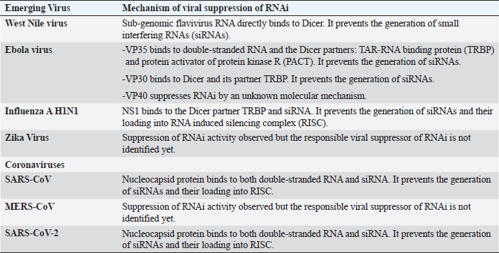
AbstractThe primary objective of this review is to describe the various mechanisms of RNA interference (RNAi) suppression in emerging RNA viruses. A search was conducted using MeSH terms such as “emerging RNA viruses,” “RNA interference,” “RNAi suppression in West Nile virus, SARS-CoV, MERS-CoV, SARS-CoV-2, Ebola virus, H1N1, and Zika virus,” “viral suppressors of RNAi in West Nile virus, SARS-CoV, MERS-CoV, SARS-CoV-2, Ebola virus, H1N1, and Zika virus,” and “siRNA prophylaxis and treatment for emerging viruses” in PubMed, Scopus, Web of Science, ScienceDirect, and Google Scholar databases. The inclusion criteria for this review encompass articles published in English between 2000 and 2023 on RNAi suppression in emerging viruses. Excluded were studies that inhibited viral replication through methods other than RNAi suppression. Viral suppressors of RNAi (VSR) typically silence RNAi by binding to viral double-stranded RNA intermediate and small interfering RNA (siRNA). Zika virus and coronaviruses execute RNAi suppression through VSR interactions with RNA. However, unique mechanisms of RNAi suppression were observed in West Nile virus (WNV), Ebola virus, and Influenza A–H1N1. In WNV, a unique protein-RNA interaction was noted, wherein subgenomic RNA directly interacts with Dicer to inhibit RNAi. In Ebola and the H1N1 virus, protein–protein interactions are employed to silence RNAi. VP35 of the Ebola virus binds to Dicer partner proteins, TAR-RNA binding protein (TRBP), and protein activator of protein kinase R (PACT), while the nonstructural protein 1 (NS1) of H1N1 binds to TRBP to suppress RNAi. Several research studies have demonstrated that by varying the delivery and dosage of siRNAs, they can be used as tools to effectively hinder the replication of emerging viruses in both cell cultures and animal models. Therefore, siRNAs can be used for prophylaxis and postexposure treatment of these viruses. Currently, no vaccines or antivirals exist for many emerging viruses, which employ diverse mechanisms to suppress RNAi. Nevertheless, siRNAs provide an attractive novel tool for prophylactic and postexposure treatment of these viruses. Keywords: Emerging RNA viruses, RNAi, RNAi suppression, Viral suppressor of RNAi. IntroductionEmerging viral diseases are diseases that have either appeared in a population for the first time or previously existed but are rapidly increasing in incidence or geographical range. Many viruses classified as emerging viruses are RNA viruses. These emerging viruses successfully replicate, mutate, adapt, and spread. The virus’s successful replication and spread primarily hinge on its capacity to circumvent the host’s and the biological vector’s antiviral defense mechanisms. The host mounts antiviral defense mechanisms through innate and acquired immunity, as well as RNA interference (RNAi) mechanisms. Innate and acquired immunity components engaged in combating viruses include phagocytes, natural killer cells, interferons (IFN), and T cytotoxic cells. IFNs play a crucial role in combating viral infections after successful viral replication in cells, while RNAi interferes with viral replication. RNAi is a highly evolved intracellular antiviral defense mechanism found in eukaryotic cells (Schuster et al., 2019; Jin et al., 2021). It is an RNA-guided gene-silencing pathway that operates at the post-transcriptional level. Central molecules in the RNAi process are microRNAs (miRNAs) and small interfering RNAs (siRNAs), playing essential roles in gene regulation and antiviral defense, respectively. During viral replication within the cell, the viral double-stranded RNA (dsRNA) intermediate is cleaved by Dicer to generate siRNAs, typically 21–25 nucleotides long. One of the siRNA strands loads into the RNA-induced silencing complex (RISC), which contains a protein called Argonaut with RNAseH activity (Wilson and Doudna, 2013). The loaded RISC with siRNA seeks and binds complementary viral RNA, subsequently cleaving it to prevent virus replication (Bagasra and Prilliman, 2004). Furthermore, viruses have evolved counter-defense mechanisms to overcome host RNAi. These viruses encode proteins with viral suppressors of RNAi (VSR) activity, which bind to dsRNA and siRNA, preventing the generation of siRNA and its binding to RISC, respectively. VSRs sequester them to suppress RNAi-mediated antiviral responses (Singh et al., 2009; Bivalkar-Mehla et al., 2011; Burgyán and Havelda, 2011; Song et al., 2011; Shaik Syed Ali et al., 2015). Some VSRs employ unique mechanisms by interacting with proteins involved in RNAi modulation, rather than directly binding with RNA. RNAi activity and its counter-suppression by VSRs have been observed in both hosts and biological vectors, resulting in unhindered virus replication. At present, there are no vaccines or antivirals for many emerging viruses. Numerous studies have shown that siRNAs remain the best approach for prophylaxis and postexposure treatment of these viruses. This review article focuses on recent research concerning the biology of VSRs in emerging RNA viruses, including West Nile virus (WNV), coronaviruses [severe acute respiratory syndrome coronavirus (SARS-CoV), Middle East respiratory syndrome coronavirus (MERS-CoV), and severe acute respiratory syndrome coronavirus 2 (SARS-CoV-2)], Influenza A-H1N1, Ebola virus, Zika virus, and the use of siRNA for prophylaxis and treatment of these viruses. Search strategy and selection criteriaA search was conducted using MeSH terms such as “emerging RNA viruses,” “RNA interference,” “RNAi suppression in West Nile virus, SARS-CoV, MERS-CoV, SARS-CoV-2, Ebola virus, H1N1, and Zika virus,” “viral suppressors of RNAi in West Nile virus, SARS-CoV, MERS-CoV, SARS-CoV-2, Ebola virus, H1N1, and Zika virus,” and “siRNA prophylaxis and treatment for emerging viruses” in PubMed, Scopus, Web of Science, ScienceDirect, and Google Scholar databases. Articles published in English between 2000 and 2023 related to RNAi suppression in emerging viruses are included. Articles focusing on inhibiting viral replication through methods other than RNAi suppression are excluded. RNAi suppressors of emerging RNA virusesRNAi silencing in WNV WNV, a flavivirus transmitted to humans through mosquito bites, is known to cause neurological diseases such as encephalitis and meningitis. The virus naturally persists in animal hosts and mosquito vectors (Ain-Najwa et al., 2020; Schvartz et al., 2020; Taieb et al., 2020). During virus replication in eukaryotic cells, VSR proteins encoded by the viral genome are produced to counteract antiviral RNAi. Among flaviviruses, dengue virus is the only virus known to encode VSRs (Qiu et al., 2020). However, investigations have revealed that during WNV replication, a noncoding RNA called sub-genomic flavivirus RNA (SfRNA) is expressed. SfRNA, approximately 525-nt in length, is derived from the 3’ untranslated region (3’UTR) and possesses a complex structure with numerous stem-loops (Markoff, 2003; Funk et al., 2010). SfRNA functions as a VSR in WNV infections by competitively binding to Dicer and inhibiting siRNA generation. This activity has been observed in both mammalian and insect cells. The precise regulatory mechanism governing SfRNA expression remains unknown, although it has been found to operate in a concentration-dependent manner (Schnettler et al., 2012). This represents a unique mechanism by which subgenomic RNA modulates RNAi by directly interacting with Dicer to prevent siRNA generation. RNAi silencing in Coronaviruses SARS-CoV, MERS-CoV, and SARS-CoV-2 caused pandemics in 2002, 2012, and 2019, respectively. All these viruses lead to acute respiratory distress syndrome. Two independent studies have shown that SARS-CoV encodes a VSR to counteract host antiviral RNAi. According to studies conducted in 2010 and 2015, the 7a accessory protein and nucleocapsid (Nc) protein were identified as the VSRs in SARS-CoV. VSR activity of SARS-CoV was demonstrated in mammalian and lung carcinoma cells using both wild-type and mutant 7a accessory proteins. The 7a accessory protein is 122 amino acids long and exhibited VSR activity independently of its N-terminal and C-terminal domains. Residues 32–89 were found to play a crucial role in VSR activity (Karjee et al., 2010). Besides its VSR activity, the 7a accessory protein also exhibits other biological activities, such as IFN antagonism, induction of apoptosis in a caspase-dependent manner, and induction of cell cycle arrest (Tan et al., 2004; Yuan et al., 2006; Kopecky-Bromberg et al., 2007). The Nc protein is a basic protein with a nonspecific affinity for RNA and DNA, playing a crucial role in SARS-CoV replication (Almazán et al., 2004; Tylor et al., 2009). Its C-terminal domain has a stronger binding affinity for RNA than the N-terminal domain (Luo et al., 2006). The Nc protein efficiently suppresses RNAi in mammalian cells triggered by short hairpin RNAs or siRNAs. In particular, residues Lys258 and Lys262 are crucial for suppressing RNAi. It accomplishes this by binding to dsRNA and siRNA and prevents the generation of siRNA and its binding to RISC, respectively (Cui et al., 2015). Although both Nc and the 7a accessory protein are VSRs in SARS-CoV, the Nc protein is a more promising VSR since it is highly conserved and encoded by all coronaviruses. Despite the efficient VSR activity of the Nc protein in SARS-CoV and SARS-CoV-2, it failed to show similar activity in MERS-CoV. Protein 4a, a dsRNA-binding IFN antagonist that inhibits protein kinase R, may have a VSR function in MERS-CoV (Niemeyer et al., 2013; Cui et al., 2015; Rabouw et al., 2016). While no studies have identified a VSR in MERS-CoV, research has shown that siRNAs have an inhibitory effect on MERS-CoV replication in cell lines (El-Kafrawy et al., 2021; Sohrab et al., 2021). The sequence homology between the Nc protein of coronaviruses is approximately 90% (Bai et al., 2021). Residues Lys258 and Lys262 are highly conserved in the Nc protein of SARS-CoV and SARS-CoV-2, and they might be crucial for interacting with RNA. The Nc protein of SARS-CoV-2 binds dsRNA and siRNA, sequestering dsRNA from Dicer and siRNA from RISC, respectively. It suppresses RNAi by preventing the generation of siRNAs and their binding to RISC (Mu et al., 2020). Other biological functions of the Nc protein include packaging of viral genome, enhancement of viral transcription and assembly. The N-terminal domain and serine-arginine-rich linker region of the Nc have high cooperativity in RNA binding affinity. Therefore, both the N-terminal and C-terminal domains play an important role in packing the RNA. In vitro studies have shown that the SARS-CoV-2 Nc protein utilizes liquid–liquid phase separation to promote the assembly of RNA-dependent RNA polymerase with polyU RNA, enabling initiation and elongation during transcription (Bai et al., 2021). The VSR activity of Nc and mutations in the spike protein of SARS-CoV-2 may have played an essential role in enhancing transmissibility and pathogenesis in SARS-CoV-2 and its variants. RNAi silencing in Ebola virus Ebola virus causes Ebola virus disease and has caused several outbreaks between 1976 and 2022. Two independent studies have shown that the Ebola virus encodes more than one VSR, including VP30, VP35, and VP40. Haasnoot et al. (2007) demonstrated that the VP35 protein has VSR activity in the Ebola virus, while Fabozzi et al. (2011) showed that, in addition to VP35, the virus encodes two more VSRs, VP30, and VP40. The VSR activity of VP35 was experimentally demonstrated by restoring Influenza A virus replication in an nonstructural protein 1 (NS1) deletion mutant, which serves as the VSR of the Influenza A virus. The dsRNA binding domain of VP35 shares sequence similarity with NS1 of Influenza A, and residues 304–314 in VP35 are crucial for binding dsRNA, especially the positively charged residues Lys309 and Arg312. In addition, VP35 directly interacts with Dicer partners, TAR-RNA binding protein (TRBP) and protein activator of protein kinase R (PACT), but not with Dicer. Mutation of residues Lys309 and Arg312 did not impact the interaction with TRBP and PACT, demonstrating their vital role only in RNA binding (Haasnoot et al., 2007). TRBP and PACT are dsRNA binding proteins that associate with Dicer for siRNA generation (Kok et al., 2007). Therefore, VP35 is one of the few VSRs capable of suppressing RNAi by preventing siRNA generation through protein–protein interactions. Another biological function of VP35 is its role as an IFN antagonist, but it can suppress RNAi independently of IFN effects. Despite having a short region of basic residues and a zinc finger motif in the N-terminal domain, VP30 of the Ebola virus did not exhibit RNA binding activity. The C-terminal domain of VP30 interacts with Dicer in the presence of siRNA and with the Dicer partner TRBP in the absence of siRNA. It suppresses RNAi by preventing siRNA generation (Fabozzi et al., 2011). Although VP40 is considered a VSR, the molecular mechanism by which it inhibits RNAi is still unknown. RNAi silencing in H1N1 H1N1 is an Influenza A virus belonging to the Orthomyxoviridae family and has been responsible for pandemics since 1918, with the most recent one occurring in 2019. NS1 has several functions, including the inhibition of IFN, viral RNA replication, and viral protein synthesis (Hale et al., 2008). NS1 modulates the host immune response by suppressing RNAi, in addition to IFN antagonism. NS1 efficiently binds to siRNAs and suppresses RNAi in both plants and mammalian cells by preventing the siRNA loading into RISC (Bucher et al., 2004; Li et al., 2016). When compared to other subtypes such as H3N2, H5N1, and H7N7, the NS1 expressed by H1N1 is particularly effective at silencing RNAi. This suggests that the ability of NS1 to suppress RNAi may vary among Influenza types and could contribute to the enhanced virulence and pathogenicity of H1N1 (de Vries et al., 2009). NS1 modulates the RNAi machinery similarly to VP35 of the Ebola virus, possibly due to their high sequence similarity. While VP35 binds both TRBP and PACT, NS1 exclusively binds to TRBP. The RNA binding domain in the N-terminus interacts with TRBP and mediates RNAi suppression by preventing siRNA generation. The residues Arg38 and Lys41 in NS1 are crucial for this interaction (Wang et al., 2022). VSRs typically silence RNAi suppression by binding to dsRNA or siRNA substrates, but NS1 of H1N1 and VP35 of Ebola are among the few VSRs discovered so far that can silence RNAi through a unique mechanism involving protein–protein interaction. RNAi silencing in Zika virus The Zika virus is an arbovirus that belongs to the flavivirus group. Although the virus has mainly remained in Africa, it has caused recent outbreaks in the Americas, Europe, and Asia. Zika virus-specific siRNAs have been found to suppress viral replication in both mosquitoes and mammals (Varjak et al., 2017; Xie and Shi, 2019). siRNAs have also been demonstrated in human neural progenitor cells and muscle tissues (Xu et al., 2019; Zhang et al., 2020). Knocking down the expression of RNAi-Dicer/Argonaut 2 components has been shown to enhance the production of siRNAs (Xu et al., 2019). Surprisingly, VSRs have not been discovered in many flaviviruses except Dengue (Qiu et al., 2020). Although no VSRs have been discovered yet in the Zika virus, there is a probability that it might encode a VSR similar to other RNA viruses or might encode sfRNA, as found in flaviviruses such as WNV, to silence RNAi. Small RNAs: A promising antiviral approach for emerging infectious diseasesFor many emerging viruses, licensed vaccines or treatments are currently unavailable. Although vaccines are in development and showing promise in animal models, the emergence of mutations in the viruses may render them ineffective. Conversely, mutations have little to no impact on antiviral small RNAs. Therefore, small RNAs represent a novel class of antiviral agents with the potential to prevent and treat emerging viral infections, particularly siRNAs, which have demonstrated promising effects in cell culture and animal models. The effectiveness of siRNAs in combating viral infections depends on the timing of RNA delivery relative to viral challenge. For example, siRNAs targeting the 3’UTR of WNV have been shown to suppress virus replication in cell culture (Anthony et al., 2009). Similar antiviral effects were observed with siRNAs targeting the NS2A and NS5 regions of WNV when administered 48 hours postinfection (Karothia et al., 2018). Another study demonstrated that miRNAs targeting the NS5 region significantly reduced WNV viral titers (Karothia et al., 2020). In animal studies, the administration of siRNAs 24 hours before the WNV challenge reduced viral loads and protected mice (Bai et al., 2005). A novel strategy involved complexing siRNAs with a neuroinvasive peptide derived from the Rabies virus, targeting the E-protein of WNV. Intranasal instillation of this complex was effective against neuroinvasive WNV disease in a mouse model and provided long-term immunity against WNV infections (Beloor et al., 2018). In the case of SARS-CoV, siRNAs targeting the spike gene, 3’ UTR, and leader sequence have been tested in cell culture and demonstrated effectiveness in inhibiting viral replication (Li et al., 2005a; Wu et al., 2005). In macaque monkeys, siRNAs targeting the spike and ORF1b showed a synergistic effect in prophylaxis and therapeutic treatments (Li et al., 2005b). Although several vaccines are available for COVID-19, the rapid emergence of variants with the potential to evade vaccine-acquired immunity makes siRNAs a valuable tool for prophylaxis and adjunctive therapy. An inhalable siRNA, C6G25S, was found to be effective against SARS-CoV-2 and its variants (alpha, delta, gamma, and epsilon) in mice. It significantly reduced virion production when used as both prophylaxis and treatment (Chang et al., 2022). Another study demonstrated the effectiveness of siRNAs targeting highly conserved regions of SARS-CoV-2 against both wildtype and relevant variants in human cells and mice (Hariharan et al., 2023). A study by Sartaj Sohrab et al. (2023) demonstrated effective prophylaxis against MERS-CoV viral infection in cell culture using siRNAs targeting ORF 1ab, although postexposure treatment experiments were not analyzed. There are very limited studies available for the Ebola virus in the context of small RNA prophylaxis and therapy. A study by O’Donnell et al. (2023) showed promising in vitro effects of artificial miRNAs in reducing viral replication but failed to protect mice in postexposure treatment. Arbutus Biopharma Pharmaceutical initiated experiments with a siRNA-based Ebola drug named TKM-Ebola, which passed Phase 1 trials in 2014 and advanced to Phase 2 trials but was abruptly ceased in 2015 (Sarisozen et al., 2015). The low number of research studies on the Ebola virus may be attributed to the challenges associated with conducting experiments, as the virus is classified under biosafety level 4 (BSL-4). BSL-4 laboratories must meet stringent requirements and obtain clearance to handle it. Table 1. Mechanisms of suppression of RNAi in emerging RNA viruses.
Current vaccines and anti-influenza drugs are only partially effective against Influenza A due to mutations and drug resistance, making prophylaxis and treatment through siRNAs a promising approach. For instance, siRNAs targeting the Nc effectively inhibit the virus in cell culture (Tompkins et al., 2004; Piasecka et al., 2020). A cocktail of siRNAs targeting various proteins of the Influenza virus, such as Nc, nonstructural proteins, and RNA-dependent RNA polymerase, has demonstrated effective antiviral effects against H1N1, H1N1pdm09, H5N2, and H7N9 (Brodskaia et al., 2018). Intranasal administration of siRNAs specifically targeting the influenza A virus Nc and acidic polymerase significantly decreased virus titers of Influenza A H5 and H7 in the lungs of mice, achieving success in both prophylaxis and therapy (Tompkins et al., 2004). Overexpression of long noncoding RNA 61 in the host impedes the multiplication of H1N1, H5N1, and H7N9. The delivery of long noncoding RNA 61 in mice restricted the multiplication of the virus by interfering with several steps, including viral entry, RNA synthesis, and release. Therefore, siRNAs and long noncoding RNAs are promising therapeutic targets for Influenza A virus infections (Hu et al., 2023). While studies on siRNAs and the Zika virus have been conducted in mosquito cell lines, demonstrating their impact on virus replication, animal studies are not yet available. While suppressing, the Dicer gene has been observed to enhance Zika virus replication, siRNAs have proven effective in inhibiting Zika virus replication, even in cases of co-infection with chikungunya and dengue viruses (Leggewie et al., 2023; Merkling et al., 2023). In summary, research studies suggest that siRNAs are a promising tool for prophylaxis and treatment of emerging viral infections, offering hope for preventing future epidemics and pandemics. ConclusionCurrently, there are no vaccines or antivirals available for emerging viruses. Emerging RNA viruses employ various mechanisms to suppress RNAi (Table 1). Nonetheless, research has shown that by increasing the dosage and exploring different delivery methods, siRNA can offer an appealing novel prophylactic and postexposure treatment option for these emerging viruses. Conflict of interestThe author has no relevant financial or nonfinancial interests to disclose. ReferencesAin-Najwa, M.Y., Yasmin, A.R., Omar, A.R., Arshad, S.S., Abu, J., Mohammed, H.O., Kumar, K., Loong, S.K., Rovie-Ryan, J.J. and Mohd-Kharip-Shah, A.K. 2020. Evidence of West Nile virus infection in migratory and resident wild birds in west coast of peninsular Malaysia. One Health 10, 100134. Almazán, F., Galán, C. and Enjuanes, L. 2004. The nucleoprotein is required for efficient coronavirus genome replication. J. Virol. 78, 12683–12688. Anthony, K.G., Bai, F., Krishnan, M.N., Fikrig, E. and Koski, R.A. 2009. Effective siRNA targeting of the 3′ untranslated region of the West Nile virus genome. Antiviral Res. 82, 166–168. Bagasra, O. and Prilliman, K.R. 2004. RNA interference: the molecular immune system. J. Mol. Histol. 35, 545–553. Bai, Z., Cao, Y., Liu, W. and Li, J. 2021. The SARS-CoV-2 nucleocapsid protein and its role in viral structure, biological functions, and a potential target for drug or vaccine mitigation. Viruses 13, 1115. Bai, F., Wang, T., Pal, U., Bao, F., Gould, L.H. and Fikrig, E. 2005. Use of RNA interference to prevent lethal murine west Nile virus infection. J. Infect. Dis. 191, 1148–1154. Beloor, J., Maes, N., Ullah, I., Uchil, P., Jackson, A., Fikrig, E., Lee, S.K. and Kumar, P. 2018. Small interfering RNA-mediated control of virus replication in the CNS is therapeutic and enables natural immunity to West Nile virus. Cell Host Microbe 23, 549–556.e3. Bivalkar-Mehla, S., Vakharia, J., Mehla, R., Abreha, M., Kanwar, J.R., Tikoo, A. and Chauhan, A. 2011. Viral RNA silencing suppressors (RSS): novel strategy of viruses to ablate the host RNA interference (RNAi) defense system. Virus Res. 155, 1–9. Brodskaia, A.V., Timin, A.S., Gorshkov, A.N., Muslimov, A.R., Bondarenko, A.B., Tarakanchikova, Y.V., Zabrodskaya, Y.A., Baranovskaya, I.L., Il’inskaja, E.V., Sakhenberg, E.I. and Sukhorukov, G.B. 2018. Inhibition of influenza A virus by mixed siRNAs, targeting the PA, NP, and NS genes, delivered by hybrid microcarriers. Antiviral Res. 158, 147–160. Bucher, E., Hemmes, H., de Haan, P., Goldbach, R. and Prins, M. 2004. The influenza A virus NS1 protein binds small interfering RNAs and suppresses RNA silencing in plants. J. Gen. Virol. 85, 983–991. Burgyán, J. and Havelda, Z. 2011. Viral suppressors of RNA silencing. Trends Plant Sci. 16, 265–272. Chang, Y.C., Yang, C.F., Chen, Y.F., Yang, C.C., Chou, Y.L., Chou, H.W., Chang, T.Y., Chao, T.L., Hsu, S.C., Ieong, S.M. and Tsai, Y.M. 2022. A siRNA targets and inhibits a broad range of SARS-CoV-2 infections including Delta variant. EMBO Mol. Med. 14, e15298. Cui, L., Wang, H., Ji, Y., Yang, J., Xu, S., Huang, X., Wang, Z., Qin, L., Tien, P., Zhou, X., Guo, D. and Chen, Y. 2015. The nucleocapsid protein of coronaviruses acts as a viral suppressor of RNA silencing in mammalian cells. J. Virol. 89, 9029–9043. de Vries, W., Haasnoot, J., Fouchier, R., de Haan, P. and Berkhout, B. 2009. Differential RNA silencing suppression activity of NS1 proteins from different influenza A virus strains. J. Gen. Virol. 90, 1916–1922. El-Kafrawy, S.A., Sohrab, S.S., Mirza, Z., Hassan, A.M., Alsaqaf, F. and Azhar, E.I. 2021. In vitro inhibitory analysis of rationally designed siRNAs against MERS-CoV replication in Huh7 cells. Molecules 26, 2610. Fabozzi, G., Nabel, C.S., Dolan, M.A. and Sullivan, N.J. 2011. Ebolavirus proteins suppress the effects of small interfering RNA by direct interaction with the mammalian RNA interference pathway. J. Virol. 85, 2512–2523. Funk, A., Truong, K., Nagasaki, T., Torres, S., Floden, N., Balmori Melian, E., Edmonds, J., Dong, H., Shi, P.Y. and Khromykh, A.A. 2010. RNA structures required for production of subgenomic flavivirus RNA. J. Virol. 84, 11407–11417. Haasnoot, J., de Vries, W., Geutjes, E.J., Prins, M., de Haan, P. and Berkhout, B. 2007. The ebola virus VP35 protein is a suppressor of RNA silencing. PLoS Pathog. 3, e86. Hale, B.G., Randall, R.E., Ortin, J. and Jackson, D. 2008. The multifunctional NS1 protein of influenza A viruses. J. Gen. Virol. 89, 2359–2376. Hariharan, V.N., Shin, M., Chang, C.W., O’Reilly, D., Biscans, A., Yamada, K., Guo, Z., Somasundaran, M., Tang, Q., Monopoli, K. and Krishnamurthy, P.M. 2023. Divalent siRNAs are bioavailable in the lung and efficiently block SARS-CoV-2 infection. Proc. Natl. Acad. Sci. U S A 120, e2219523120. Hu, J., Zhang, L., Zheng, X., Wang, G., Chen, X., Hu, Z., Chen, Y., Wang, X., Gu, M., Hu, S. and Liu, X. 2023. Long noncoding RNA #61 exerts a broad anti-influenza a virus effect by its long arm rings. Antiviral Res. 215, 105637. Jin, Y., Zhao, J.H. and Guo, H.S. 2021. Recent advances in understanding plant antiviral RNAi and viral suppressors of RNAi. Curr. Opin. Virol. 46, 65–72. Karjee, S., Minhas, A., Sood, V., Ponia, S.S., Banerjea, A.C., Chow, V.T., Mukherjee, S.K. and Lal, S.K. 2010. The 7a accessory protein of severe acute respiratory syndrome coronavirus acts as an RNA silencing suppressor. J. Virol. 84, 10395–10401. Karothia, D., Dash, P.K., Parida, M., Bhagyawant, S. and Kumar, J.S. 2018. Inhibition of West Nile virus replication by bifunctional siRNA targeting the NS2A and NS5 conserved region. Curr. Gene Ther. 18, 180–190. Karothia, D., Dash, P.K., Parida, M., Bhagyawant, S.S. and Kumar, J.S. 2020. Vector derived artificial miRNA mediated inhibition of West Nile virus replication and protein expression. Gene 729, 144300. Kok, K.H., Ng, M.H.J., Ching, Y.P. and Jin, D.Y. 2007. Human TRBP and PACT directly interact with each other and associate with dicer to facilitate the production of small interfering RNA. J. Biol. Chem. 282, 17649–17657. Kopecky-Bromberg, S.A., Martínez-Sobrido, L., Frieman, M., Baric, R.A. and Palese, P. 2007. Severe acute respiratory syndrome coronavirus open reading frame (ORF) 3b, ORF 6, and nucleocapsid proteins function as interferon antagonists. J. Virol. 81, 548–557. Leggewie, M., Scherer, C., Altinli, M., Gestuveo, R.J., Sreenu, V.B., Fuss, J., Vazeille, M., Mousson, L., Badusche, M., Kohl, A. and Failloux, A.B. 2023. The Aedes aegypti RNA interference response against Zika virus in the context of co-infection with dengue and chikungunya viruses. PLoS Negl. Trop. Dis. 17, e0011456. Li, T., Zhang, Y., Fu, L., Yu, C., Li, X., Li, Y., Zhang, X., Rong, Z., Wang, Y., Ning, H. and Liang, R. 2005a. siRNA targeting the leader sequence of SARS-CoV inhibits virus replication. Gene Ther. 12, 751–761. Li, B.J., Tang, Q., Cheng, D., Qin, C., Xie, F.Y., Wei, Q., Xu, J., Liu, Y., Zheng, B.J., Woodle, M.C. and Zhong, N. 2005b. Using siRNA in prophylactic and therapeutic regimens against SARS coronavirus in Rhesus macaque. Nat. Med. 11, 944–951. Li, Y., Basavappa, M., Lu, J., Dong, S., Cronkite, D.A., Prior, J.T., Reinecker, H.C., Hertzog, P., Han, Y., Li, W.X. and Cheloufi, S. 2016. Induction and suppression of antiviral RNA interference by influenza A virus in mammalian cells. Nat. Microbiol. 2, 16250. Luo, H., Chen, J., Chen, K., Shen, X. and Jiang, H. 2006. Carboxyl terminus of severe acute respiratory syndrome coronavirus nucleocapsid protein: self-association analysis and nucleic acid binding characterization. Biochemistry 45, 11827–11835. Markoff, L. 2003. 5′- and 3′-noncoding regions in flavivirus RNA. Adv. Virus. Res. 59, 177–228. Merkling, S.H., Crist, A.B., Henrion-Lacritick, A., Frangeul, L., Couderc, E., Gausson, V., Blanc, H., Bergman, A., Baidaliuk, A., Romoli, O. and Saleh, M.C. 2023. Multifaceted contributions of Dicer2 to arbovirus transmission by Aedes aegypti. Cell Rep. 42, 112977. Mu, J., Xu, J., Zhang, L., Shu, T., Wu, D., Huang, M., Ren, Y., Li, X., Geng, Q., Xu, Y., Qiu, Y. and Zhou, X. 2020. SARS-CoV-2-encoded nucleocapsid protein acts as a viral suppressor of RNA interference in cells. Sci. China Life Sci. 63, 1413–1416. Niemeyer, D., Zillinger, T., Muth, D., Zielecki, F., Horvath, G., Suliman, T., Barchet, W., Weber, F., Drosten, C. and Müller, M.A. 2013. Middle east respiratory syndrome coronavirus accessory protein 4a is a type I interferon antagonist. J. Virol. 87, 12489–12495. O’Donnell, K.L., Callison, J., Feldmann, H., Hoenen, T. and Marzi, A. 2023. Single-dose treatment with VSV-EBOV expressing Ebola virus-specific artificial miRNAs does not protect mice from lethal disease. J. Infect. Dis. jiad121. Piasecka, J., Lenartowicz, E., Soszynska-Jozwiak, M., Szutkowska, B., Kierzek, R. and Kierzek, E. 2020. RNA secondary structure motifs of the Influenza A virus as targets for siRNA-mediated RNA interference. Mol. Ther. Nucleic Acids 19, 627–642. Qiu, Y., Xu, Y.P., Wang, M., Miao, M., Zhou, H., Xu, J., Kong, J., Zheng, D., Li, R.T., Zhang, R.R., Guo, Y., Li, X.F., Cui, J., Qin, C.F. and Zhou, X. 2020. Flavivirus induces and antagonizes antiviral RNA interference in both mammals and mosquitoes. Sci. Adv. 6, eaax7989. Rabouw, H.H., Langereis, M.A., Knaap, R.C., Dalebout, T.J., Canton, J., Sola, I., Enjuanes, L., Bredenbeek, P.J., Kikkert, M., de Groot, R.J. and van Kuppeveld, F.J. 2016. Middle east respiratory coronavirus accessory protein 4a inhibits PKR-mediated antiviral stress responses. PLoS Pathog. 12, e1005982. Sarisozen, C., Salzano, G. and Torchilin, V.P. 2015. Recent advances in siRNA delivery. Biomol. Concepts 6, 321–341. Sartaj Sohrab, S., Aly El-Kafrawy, S., Mirza, Z., Hassan, A.M., Alsaqaf, F. and Ibraheem Azhar, E. 2023. Delivery of siRNAs against MERS-CoV in Vero and HEK-293 cells: a comparative evaluation of transfection reagents. J. King Saud Univ. Sci. 35, 102540. Schnettler, E., Sterken, M.G., Leung, J.Y., Metz, S.W., Geertsema, C., Goldbach, R.W., Vlak, J.M., Kohl, A., Khromykh, A.A. and Pijlman, G.P. 2012. Noncoding flavivirus RNA displays RNA interference suppressor activity in insect and mammalian cells. J. Virol. 86, 13486–13500. Schuster, S., Miesen, P. and van Rij, R.P. 2019. Antiviral RNAi in insects and mammals: parallels and differences. Viruses 11, 448. Schvartz, G., Farnoushi, Y., Berkowitz, A., Edery, N., Hahn, S., Steinman, A., Lublin, A. and Erster, O. 2020. Molecular characterization of the re-emerging West Nile virus in avian species and equids in Israel, 2018, and pathological description of the disease. Parasites Vectors 13, 528. Shaik Syed Ali, P., John, J., Selvaraj, M., Kek, T.L. and Salleh, M.Z. 2015. Nodamura virus B2 amino terminal domain sensitivity to small interfering RNA. Microbiol. Immunol. 59, 299–304. Singh, G., Popli, S., Hari, Y., Malhotra, P., Mukherjee, S. and Bhatnagar, R.K. 2009. Suppression of RNA silencing by flock house virus B2 protein is mediated through its interaction with the PAZ domain of Dicer. FASEB J. 23, 1845–1857. Sohrab, S.S., El-Kafrawy, S.A., Mirza, Z., Hassan, A.M., Alsaqaf, F. and Azhar, E.I. 2021. Designing and evaluation of MERS-CoV siRNAs in HEK-293 cell line. J. Infect. Public Health 14, 238–243. Song, L., Gao, S., Jiang, W., Chen, S., Liu, Y., Zhou, L. and Huang, W. 2011. Silencing suppressors: viral weapons for countering host cell defenses. Protein Cell 2, 273–281. Tan, Y.J., Fielding, B.C., Goh, P.Y., Shen, S., Tan, T.H., Lim, S.G. and Hong, W. 2004. Overexpression of 7a, a protein specifically encoded by the severe acute respiratory syndrome coronavirus, induces apoptosis via a Caspase-dependent pathway. J. Virol. 346, 74–85. Taieb, L., Ludwig, A., Ogden, N.H., Lindsay, R.L., Iranpour, M., Gagnon, C.A., and Bicout, D.J. 2020. Bird species involved in west Nile virus epidemiological cycle in Southern Québec. Int. J. Environ. Res. Public Health 17, 4517. Tompkins, S.M., Lo, C.-Y., Tumpey, T.M. and Epstein, S.L. 2004. Protection against lethal influenza virus challenge by RNA interference in vivo. Proc. Natl. Acad. Sci. U S A 101, 8682–8686. Tylor, S., Andonov, A., Cutts, T., Cao, J., Grudesky, E., Van Domselaar, G., Li, X. and He, R. 2009. The SR-rich motif in SARS-CoV nucleocapsid protein is important for virus replication. Can. J. Microbiol. 55, 254–260. Varjak, M., Donald, C.L., Mottram, T.J., Sreenu, V.B., Merits, A., Maringer, K., Schnettler, E. and Kohl, A. 2017. Characterization of the Zika virus induced small RNA response in Aedes aegypti cells. PLoS Negl. Trop. Dis. 11, e0006010. Wang, Q., Wang, J., Xu, Y., Li, Z., Wang, B. and Li, Y. 2022. The interaction of influenza A NS1 and cellular TRBP protein modulates the function of RNA interference machinery. Front. Microbiol. 13, 859420. Wilson, R.C. and Doudna, J.A. 2013. Molecular mechanisms of RNA interference. Annu. Rev. Biophys. 42, 217–239. Wu, C.J., Huang, H.W., Liu, C.Y., Hong, C.F. and Chan, Y.L. 2005. Inhibition of SARS-CoV replication by siRNA. Antiviral Res. 65, 45–48. Xie, X. and Shi, P.Y. 2019. Anti-Zika virus RNAi in neural progenitor cells. Cell. Res. 29, 261–262. Xu, Y.P., Qiu, Y., Zhang, B., Chen, G., Chen, Q., Wang, M., Mo, F., Xu, J., Wu, J., Zhang, R.R. and Cheng, M.L. 2019. Zika virus infection induces RNAi-mediated antiviral immunity in human neural progenitors and brain organoids. Cell. Res. 29, 265–273. Yuan, X., Wu, J., Shan, Y., Yao, Z., Dong, B., Chen, B., Zhao, Z., Wang, S., Chen, J. and Cong, Y. 2006. SARS coronavirus 7a protein blocks cell cycle progression at G0/G1 phase via the cyclin D3/pRb pathway. Virology 346, 74–85. Zhang, Y., Li, Z., Ye, Z., Xu, Y., Wang, B., Wang, C., Dai, Y., Lu, J., Lu, B., Zhang, W. and Li, Y. 2020. The activation of antiviral RNA interference not only exists in neural progenitor cells but also in somatic cells in mammals. Emerg. Microbes Infect. 9, 1580–1589. | ||
| How to Cite this Article |
| Pubmed Style P. Shaik Syed Ali. Suppressors of silencers: Exploring viral suppression of RNAi in emerging RNA viruses. J Microbiol Infect Dis. 2023; 13(3): 110-117. doi:10.5455/JMID.2023.v13.i3.2 Web Style P. Shaik Syed Ali. Suppressors of silencers: Exploring viral suppression of RNAi in emerging RNA viruses. https://www.jmidonline.org/?mno=152773 [Access: May 08, 2024]. doi:10.5455/JMID.2023.v13.i3.2 AMA (American Medical Association) Style P. Shaik Syed Ali. Suppressors of silencers: Exploring viral suppression of RNAi in emerging RNA viruses. J Microbiol Infect Dis. 2023; 13(3): 110-117. doi:10.5455/JMID.2023.v13.i3.2 Vancouver/ICMJE Style P. Shaik Syed Ali. Suppressors of silencers: Exploring viral suppression of RNAi in emerging RNA viruses. J Microbiol Infect Dis. (2023), [cited May 08, 2024]; 13(3): 110-117. doi:10.5455/JMID.2023.v13.i3.2 Harvard Style P. Shaik Syed Ali (2023) Suppressors of silencers: Exploring viral suppression of RNAi in emerging RNA viruses. J Microbiol Infect Dis, 13 (3), 110-117. doi:10.5455/JMID.2023.v13.i3.2 Turabian Style P. Shaik Syed Ali. 2023. Suppressors of silencers: Exploring viral suppression of RNAi in emerging RNA viruses. Journal of Microbiology and Infectious Diseases, 13 (3), 110-117. doi:10.5455/JMID.2023.v13.i3.2 Chicago Style P. Shaik Syed Ali. "Suppressors of silencers: Exploring viral suppression of RNAi in emerging RNA viruses." Journal of Microbiology and Infectious Diseases 13 (2023), 110-117. doi:10.5455/JMID.2023.v13.i3.2 MLA (The Modern Language Association) Style P. Shaik Syed Ali. "Suppressors of silencers: Exploring viral suppression of RNAi in emerging RNA viruses." Journal of Microbiology and Infectious Diseases 13.3 (2023), 110-117. Print. doi:10.5455/JMID.2023.v13.i3.2 APA (American Psychological Association) Style P. Shaik Syed Ali (2023) Suppressors of silencers: Exploring viral suppression of RNAi in emerging RNA viruses. Journal of Microbiology and Infectious Diseases, 13 (3), 110-117. doi:10.5455/JMID.2023.v13.i3.2 |

