


| Case Report | ||
J. Microbiol. Infect. Dis., (2023), Vol. 13(2): 90–92 Case Report A middle-aged man with a mass in the brain and heartGuive Sharifi1, Abdolreza Babamahmoodi2, Shahram Sabeti3, Mohammad Hallajnejad1 and Ilad Alavi Darazam2,4*1Department of Neurosurgery, Loghman Hospital, Shahid Beheshti University of Medical Sciences, Tehran, Iran 2Infectious Diseases and Tropical Medicine Research Center, Shahid Beheshti University of Medical Sciences, Tehran, Iran 3Pathology ward, Loghman Hakim Hospital, Shahid Beheshti University of Medical Sciences, Tehran, Iran 4Infectious Diseases and Tropical Medicine, Loghman Hakim Hospital, Shahid Beheshti University of Medical Sciences, Tehran, Iran *Corresponding Author: Ilad Alavi Darazam. Infectious Diseases and Tropical Medicine, Loghman Hakim Hospital, Shahid Beheshti University of Medical Sciences, Tehran, Iran. Email: ilad13 [at] yahoo.com Submitted: 05/02/2023 Accepted: 04/06/2023, Published: 30/06/2023 © 2023 Journal of Microbiology and Infectious Diseases
AbstractBackground: Echinococcosis is a parasitic and zoonotic disease, usually caused by Echinococcus granulosus or Echinococcus multilocularis, and threatens the health and development of human societies. Worldwide, 3.6 million disability adjusted life years (DALYs) may be lost due to echinococcosis. Case Description: In this report, a 44-year-old man was presented, whose first symptoms were psychological and behavioral changes and headache. After investigations, it was found that he had a mass in the frontal part of the brain, in the heart, and left kidney and spleen without any symptoms and involvement in the liver. After surgery to remove a mass in the brain, a pathologic study confirmed that the patient had echinococcosis. After necessary radiological imaging, the patient underwent surgery to remove cysts. The cyst was successfully removed and the patient was started on albendazole. The patient was discharged in good general condition and followed up for 6 months, during which time their general condition remained very good. Conclusion: To properly diagnose hydatid cyst disease, a thorough examination of all possible sites of involvement is necessary, even if the patient has no symptoms. Keywords: Echinococcosis, Hydatid cyst, Brain mass. IntroductionEchinococcosis is a parasitic disease common to humans and animals, usually caused by tapeworms of the genus Echinococcus granulosus or Echinococcus multilocularis, and threatens the health and development of human societies. the disease is distributed worldwide and is found in all continents except Antarctica. Echinococcosis is usually found in areas where livestock farming is common and large numbers of sheep and cattle are raised (Parandin et al., 2021). the extensive movement of livestock in the world and travel between countries has turned this situation into a serious global health problem. According to the World Health Organization, more than one million people are affected by hydatid disease at any time (Dong et al., 2022). In two regions, the Middle East/North Africa and Central Asia/China, the disease occurs endemically. Worldwide, 3.6 million DALYs may be lost due to echinococcosis (da Silva, 2010). Echinococcus parasites can live in any part of the body, but they are more interested in the liver, lungs, brain, and stomach of humans. When this parasite attaches to the human body, the health condition gradually worsens. This zoonotic disease is endemic in pastoral areas around the world, where it is often spread by herders who use the viscera of infected ruminants to feed their dogs. In alveolar echinococcosis, in addition to the main life cycle in wild dogs, a life cycle has also been established in domestic dogs, which is one of the main factors of disease transmission in some regions of the world (Baumann et al., 2019). Compared to E. granulosus, the potential risk factors for E. multilocularis are more complex because its life cycle includes small mammals (mainly rodents) as intermediate hosts and canids as final hosts (Eckert and Thompson, 2017). Case DetailsA 44-year-old rancher was referred to a neurologist with complaints of mood swings, restlessness, and headaches that worsened after lifting heavy weights. The patient did not have any significant illness in their previous history and did not complain of vomiting, weight loss, or fever in their new illness. The neurological examination did not show any findings in favor of cerebral palsy or focal neurological symptoms. A CT-scan of the brain revealed an abnormal low-density mass in the right frontoparietal lobe, leading to the diagnosis of a possible cyst or abscess (Fig. 1). Antibiotic treatment was started for the patient, but after eleven days, the patient’s symptoms did not improve and even worsened. After re-imaging, the neurologist recommended that the patient see a neurosurgeon. During pre-operative investigations, consultation with a cardiologist was requested. Echocardiography revealed a moving mass confined to the anterolateral papillary muscle in the patient’s heart, even though the patient had no complaints or symptoms of heart disease (Fig. 2). Although the patient had only presented with symptoms of central nervous system involvement at the beginning of their visit, their cardiac findings were diagnosed completely by chance (a cyst confined to the anterolateral papillary muscle without any cardiovascular symptoms). In abdominal imaging, one round cyst in the spleen and upper pole of the left kidney was diagnosed. After surgery to remove the cyst from the head and subsequent follow-ups, lesions were discovered in the patient’s liver that were not present in the initial ultrasounds. The patient recovered with surgery and drug treatment with albendazole. DiscussionIn three-quarters of cases, the liver is affected, then the lungs are affected in one-fifth of cases, and other organs are affected with different frequencies. Single-organ involvement occurs in 85%–90% of cases, and a single cyst is seen in 70% of cases (Ahmed et al., 2022). Finding this disease only in the heart and brain is rare, with a frequency of 1%–2% for the brain and 0.5%–2% for the heart in all cases with hydatid cyst disease reported. The clinical presentation of cardiac hydatid cysts is uncommon and varies from completely asymptomatic forms to cases presenting severe heart failure. Arrhythmias caused by cardiac hydatid cysts have a high mortality rate but are very rare (Lu et al., 2019). Hydatid cysts in the brain are usually single, extraterritorial, and intraparenchymal, and these lesions do not cause serious symptoms until they reach large dimensions. The location of the lesion and its size are effective in clinical symptoms, and headache and throwing up are the most common symptoms observed (Svrckova et al., 2019). Psychiatric disorders such as behavioral disorders, aggression, and delusions are also reported in the articles (Asri et al., 2007).
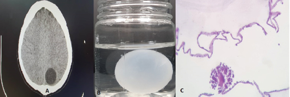
Fig. 1. (A) Brain CT-scan before the operation. (B) Mass that was removed from the frontal lobe of the brain after surgery. (C) Hydatid cyst wall with budding scolex.
Fig. 2. (A) Echocardiography of patient. (B) Chest MRI shows a mass in the heart. Obtaining a detailed history, epidemiological information, clinical symptoms of the patient, serological tests, and any morphological lesions discovered by ultrasound, CT, and MRI are the main tools for diagnosing this disease (Nabah et al., 2012). In the latest articles published in prestigious international journals, this disease is still one of the causes of problems in the central nervous system, and it requires careful attention from the health system staff (Ju and Liu, 2023; Sumit and Sunil, 2023). ConclusionConsidering a large number of patients and the effects of echinococcosis, it can be said that this disease is neglected and less reported because it takes a long time for the clinical symptoms of this disease to appear, and on the other hand, reservoirs of infection in domestic animals and wildlife have caused It is difficult to manage from the aspect of public health. Also, we strongly recommend for properly diagnose hydatid cyst disease, a thorough examination of all possible sites of involvement is necessary, even if the patient has no symptoms ReferencesAhmed, S.K., Essa, R.A. and Bapir, D.H. 2022. Uniportal video-assisted thoracoscopic surgery (u-vats) for management of pulmonary hydatid cyst: a systematic review. Ann. Med. Surg. 75, 103474; https://doi.org/10.1016/j.amsu.2022.103474 Asri, F., Tazi, I., Maaroufi, K., El Moudden, A., Ghannane, H. and Ait Benali, S. 2007. Cerebral hydatic cyst and psychiatric disorders. Two cases. L’ Encephale 33(2), 216–219; https://doi.org/10.1016/s0013-7006(07)91553-5 Baumann, S., Shi, R., Liu, W., Bao, H., Schmidberger, J., Kratzer, W. and Li, W. 2019. Worldwide literature on epidemiology of human alveolar echinococcosis: a systematic review of research published in the twenty-first century. Infection 47(5), 703–727; https://doi.org/10.1007/s15010-019-01325-2 Dong, Z., Yusup, M., Lu, Y. and Tang, B. 2022. Hydatid cyst of the heart as a rare cause of arrhythmia: a case report and review of published reports. Heart. Rhythm. Case Rep. 6, 458–462; https://doi.org/10.1016/j.hrcr.2022.04.004 da Silva, A.M. 2010. Human echinococcosis: a neglected disease. Gastroenterol. Res. Pract. 2010, 583297; https://doi.org/10.1155/2010/583297 Eckert, J. and Thompson, R.C.A. 2017. Historical aspects of echinococcosis. Adv. Parasitol. 95, 1–64; https://doi.org/10.1016/bs.apar.2016.07.003 Ju, H. and Liu, C. 2023. Cerebral alveolar echinococcosis. N. Engl. J. Med. 388(5), 453; https://doi.org/10.1056/NEJMicm2202196 Lu, Y.M., Zhang, L., Xing, Q., Zhou, X.H., Li, Y.D, Zhang, J.H, Zukela, T. and Tang, B.P. 2019. Ventricular tachycardia as the initial symptom of cardiac hydatidosis. Chin. Med. J. 132(22), 2765–2766; https://doi.org/10.1097/CM9.0000000000000520 Nabah, K., Mezzoug, N., Oufdou, H., Abrini, J. and Rharrabe, K. 2012. Epidemiological profile of echinococcosis in Morocco from 1980 to 2008. Ann. Biol. Clin. 70 (4), 457–461; https://doi.org/10.1684/abc.2012.0727 Parandin, F., Heydarpour, F., Mohebali, M., Hanafi-Bojd, A.A., Sari, A.A., Zeynali, M., Alizadeh, A., Nazari, N., Kaveh F. and Rokni, M.B. 2021. Estimation of burden of cystic Echinococcosis in Iran using disability adjusted life years (DALYs) in 2018. Iran. J. Public Health 50, 2302–2308; https://doi.org/10.18502/ijph.v50i11.7586 Svrckova, P., Nabarro, L., Chiodini, P.L. and Jäger, H.R. 2019. Disseminated cerebral hydatid disease (multiple intracranial Echinococcosis). Pract. Neurol. 19(2), 156–163; https://doi.org/10.1136/practneurol-2018-001954 Sumit, T. and Sunil, A. 2023. Cerebral cystic Echinococcosis. N. Engl. J. Med. 388(5), e10; https://doi.org/10.1056/NEJMicm2208104 | ||

| How to Cite this Article |
| Pubmed Style Sharifi G, Babamahmoodi A, Sabeti S, Hallajnejad M, Darazam IA. A middle-aged man with a mass in the brain and heart. J Microbiol Infect Dis. 2023; 13(2): 90-92. doi:10.5455/JMID.2023.v13.i2.7 Web Style Sharifi G, Babamahmoodi A, Sabeti S, Hallajnejad M, Darazam IA. A middle-aged man with a mass in the brain and heart. https://www.jmidonline.org/?mno=302657428 [Access: July 05, 2025]. doi:10.5455/JMID.2023.v13.i2.7 AMA (American Medical Association) Style Sharifi G, Babamahmoodi A, Sabeti S, Hallajnejad M, Darazam IA. A middle-aged man with a mass in the brain and heart. J Microbiol Infect Dis. 2023; 13(2): 90-92. doi:10.5455/JMID.2023.v13.i2.7 Vancouver/ICMJE Style Sharifi G, Babamahmoodi A, Sabeti S, Hallajnejad M, Darazam IA. A middle-aged man with a mass in the brain and heart. J Microbiol Infect Dis. (2023), [cited July 05, 2025]; 13(2): 90-92. doi:10.5455/JMID.2023.v13.i2.7 Harvard Style Sharifi, G., Babamahmoodi, . A., Sabeti, . S., Hallajnejad, . M. & Darazam, . I. A. (2023) A middle-aged man with a mass in the brain and heart. J Microbiol Infect Dis, 13 (2), 90-92. doi:10.5455/JMID.2023.v13.i2.7 Turabian Style Sharifi, Guive, Abdolreza Babamahmoodi, Shahram Sabeti, Mohammad Hallajnejad, and Ilad Alavi Darazam. 2023. A middle-aged man with a mass in the brain and heart. Journal of Microbiology and Infectious Diseases, 13 (2), 90-92. doi:10.5455/JMID.2023.v13.i2.7 Chicago Style Sharifi, Guive, Abdolreza Babamahmoodi, Shahram Sabeti, Mohammad Hallajnejad, and Ilad Alavi Darazam. "A middle-aged man with a mass in the brain and heart." Journal of Microbiology and Infectious Diseases 13 (2023), 90-92. doi:10.5455/JMID.2023.v13.i2.7 MLA (The Modern Language Association) Style Sharifi, Guive, Abdolreza Babamahmoodi, Shahram Sabeti, Mohammad Hallajnejad, and Ilad Alavi Darazam. "A middle-aged man with a mass in the brain and heart." Journal of Microbiology and Infectious Diseases 13.2 (2023), 90-92. Print. doi:10.5455/JMID.2023.v13.i2.7 APA (American Psychological Association) Style Sharifi, G., Babamahmoodi, . A., Sabeti, . S., Hallajnejad, . M. & Darazam, . I. A. (2023) A middle-aged man with a mass in the brain and heart. Journal of Microbiology and Infectious Diseases, 13 (2), 90-92. doi:10.5455/JMID.2023.v13.i2.7 |

